Dr. Shikhara Vijayan
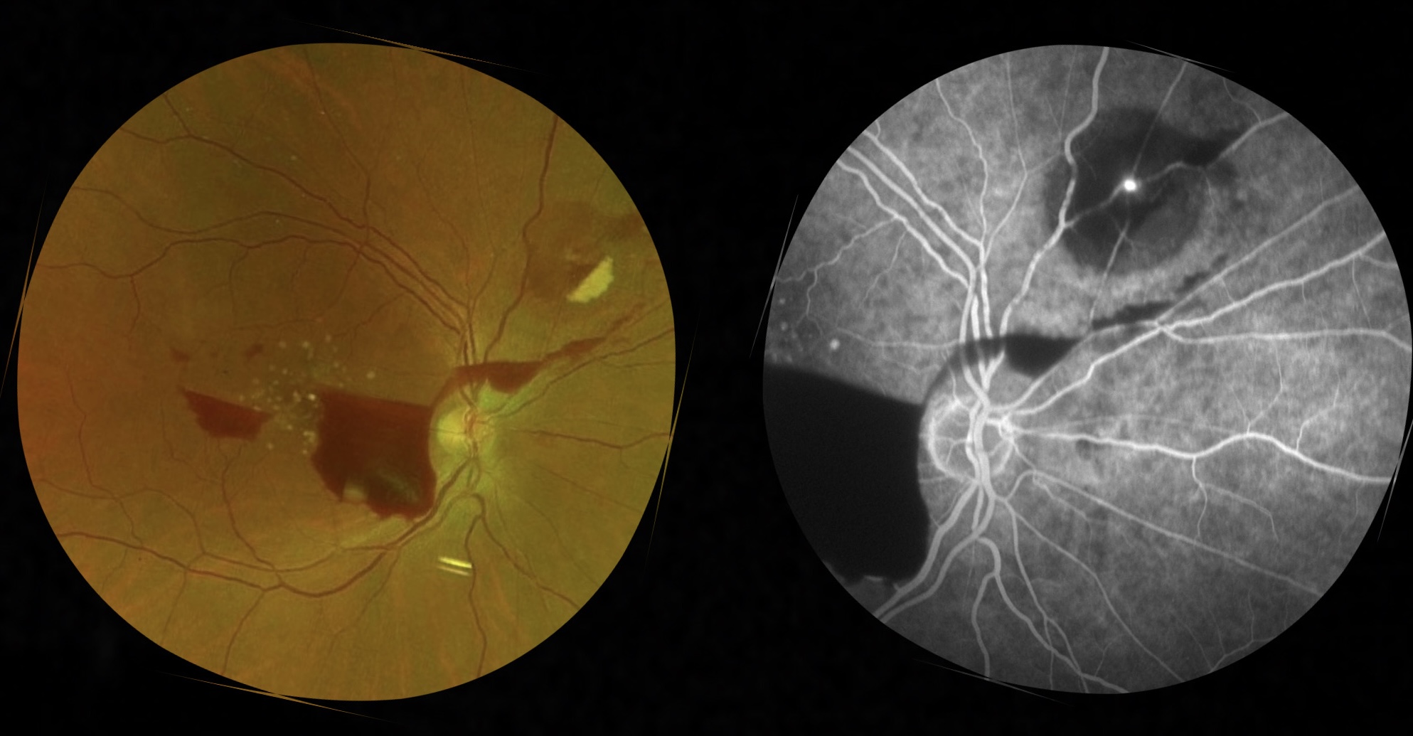
Title:The Pouring RAM
Description:This is the fundus image and fluorescein angiogram image of the right eye of a 58 year old male with a retinal artery macroaneurysm in the superonasal quadrant with multilayered hemmorhage pouring from the superonasal lesion down to the temporal retina . There are also accompanying drusens in the macular area in the same eye.
Dr. M. Nivedita
Title: A Not-so-sparkling Christmas Tree within the retina
Description: A 69 year old diabetic male presented with macular branch retinal vein occlusion with CME in the left eye. His BCVA was 6/24 OS. OCT of the left eye shows CME with intracystic hyperreflective material (ICHRM). Following intravitreal injection of ranibizumab, edema decreased and BCVA improved to 6/18.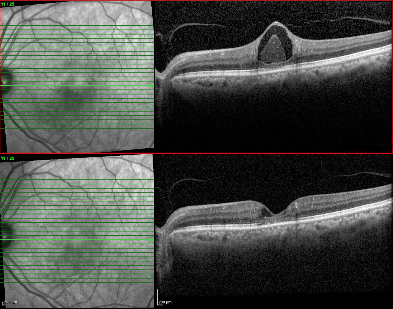
Dr. Anisha Tresa Augustin
Title: Speckled sky under a vascular storm
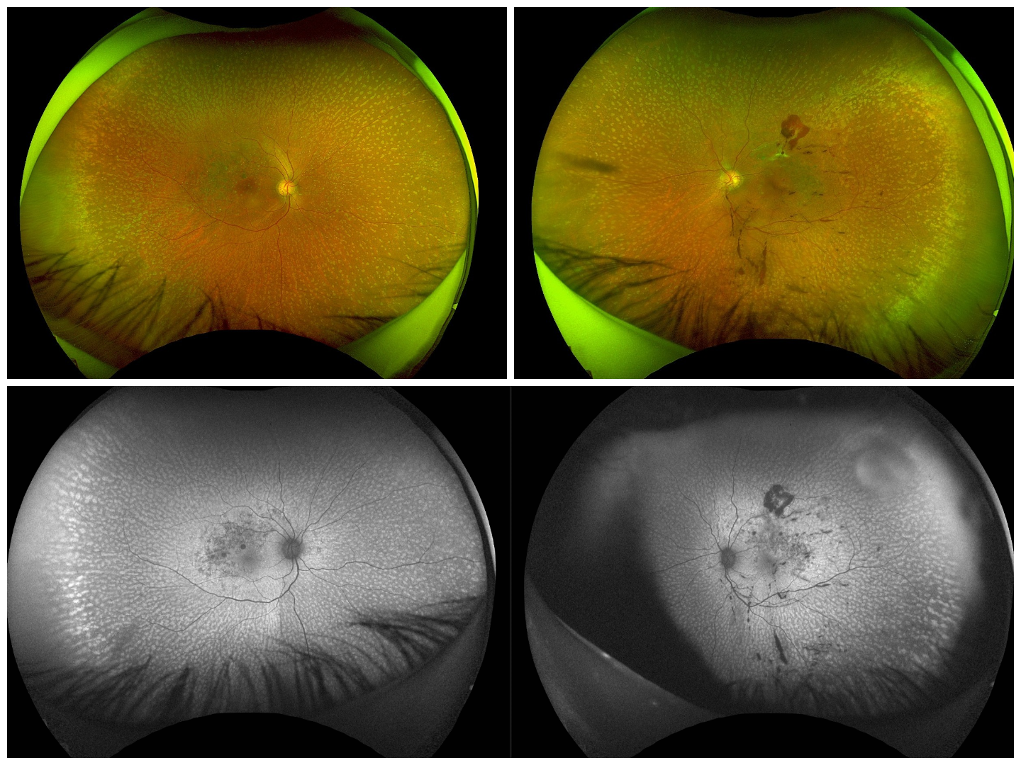
Description: A 65 year old male K/C/O Diabetes and hypertension presented with complaints of sudden onset of floaters in his left eye of 2 weeks duration. BCVA was 6/6 ,N6 in both eyes and anterior segment was normal bilaterally. Fundus examination revealed multiple whitish flecks at the mid periphery sparing the fovea in both eyes s/o fundus albipunctatus. Left eye showed a sclerosed vessel superotemporally with new vessels and dense vitreous floaters consistent with an old superotemporal branch retinal vein occlusion with neovascularization (NVE) and vitreous hemorrhage. Wide field fundus photo, fundus autofluorescence and FFA confirmed the above findings.
This is a rare case of an inherited retinal disorder which was unmasked by a vascular occlusion. It highlights the importance of multimodal imaging in delineating the features of retinal dystrophy and a superimposed vascular event, enabling accurate diagnosis and timely management.