Dr. Sherina Thomas
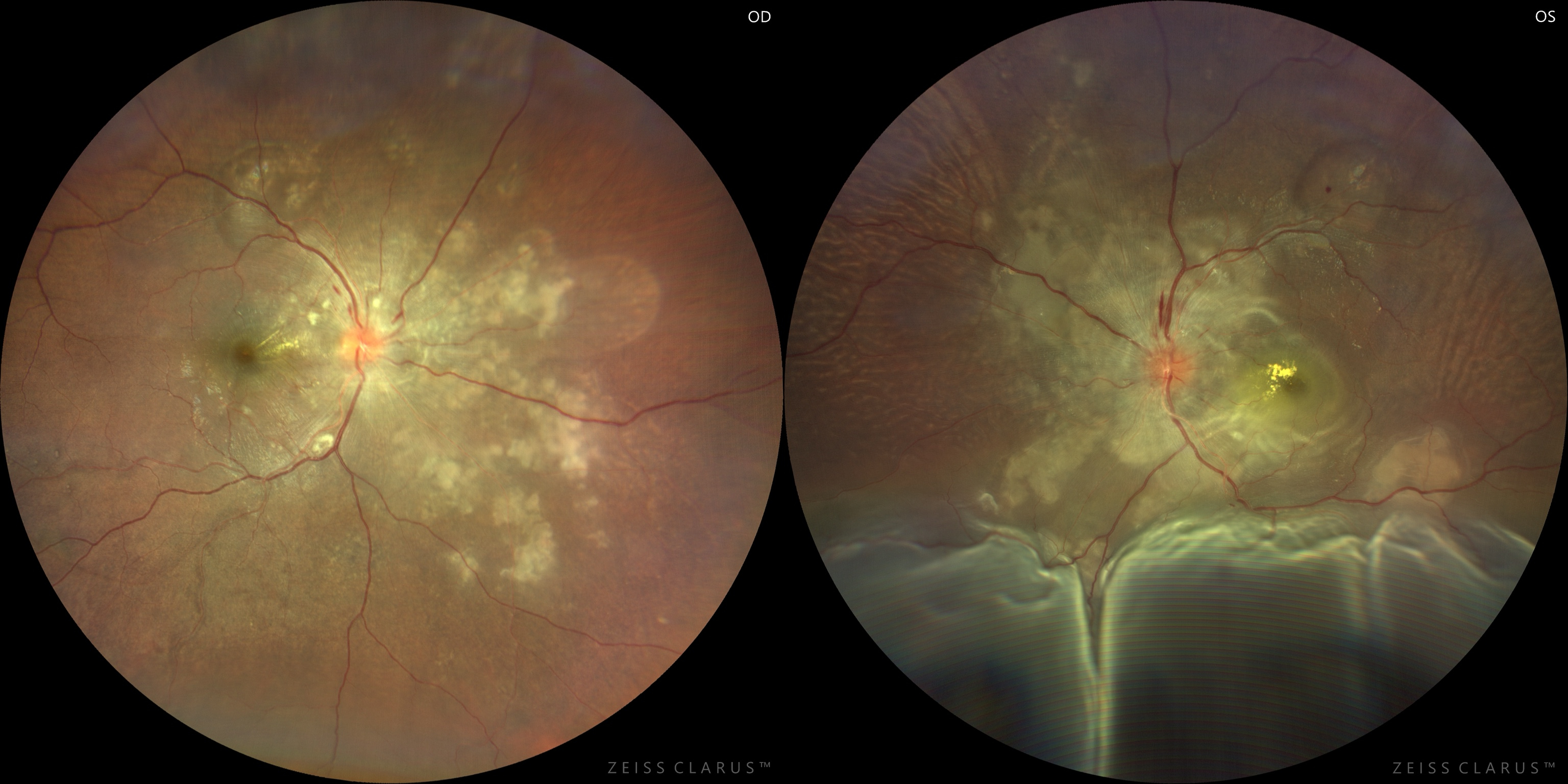
Title: Vascular Storm in the Retina
Description: A 26-year-old male presented with acute visual deterioration and headache with a blood pressure of 220/180 mmHg. Fundus imaging revealed bilateral Grade 4 hypertensive retinopathy, with extensive yellow-white lesions consistent with Elschnig spots involving the posterior pole, more prominent in the left eye. Both eyes additionally demonstrated multiple pockets of exudative retinal detachment.
These striking fundus findings illustrate the dual insult of severe hypertension on the retinal and choroidal vasculature. While retinal arteriolar injury and breakdown of the blood–retinal barrier account for the features of hypertensive retinopathy, choriocapillaris ischemia leads to retinal pigment epithelium dysfunction, Elschnig spots and serous retinal detachment. This case underscores the sight-threatening ocular consequences of a hypertensive emergency and highlights the importance of prompt recognition and systemic blood pressure control to preserve visual function.
Dr. Shraddha Raj Shrivastava
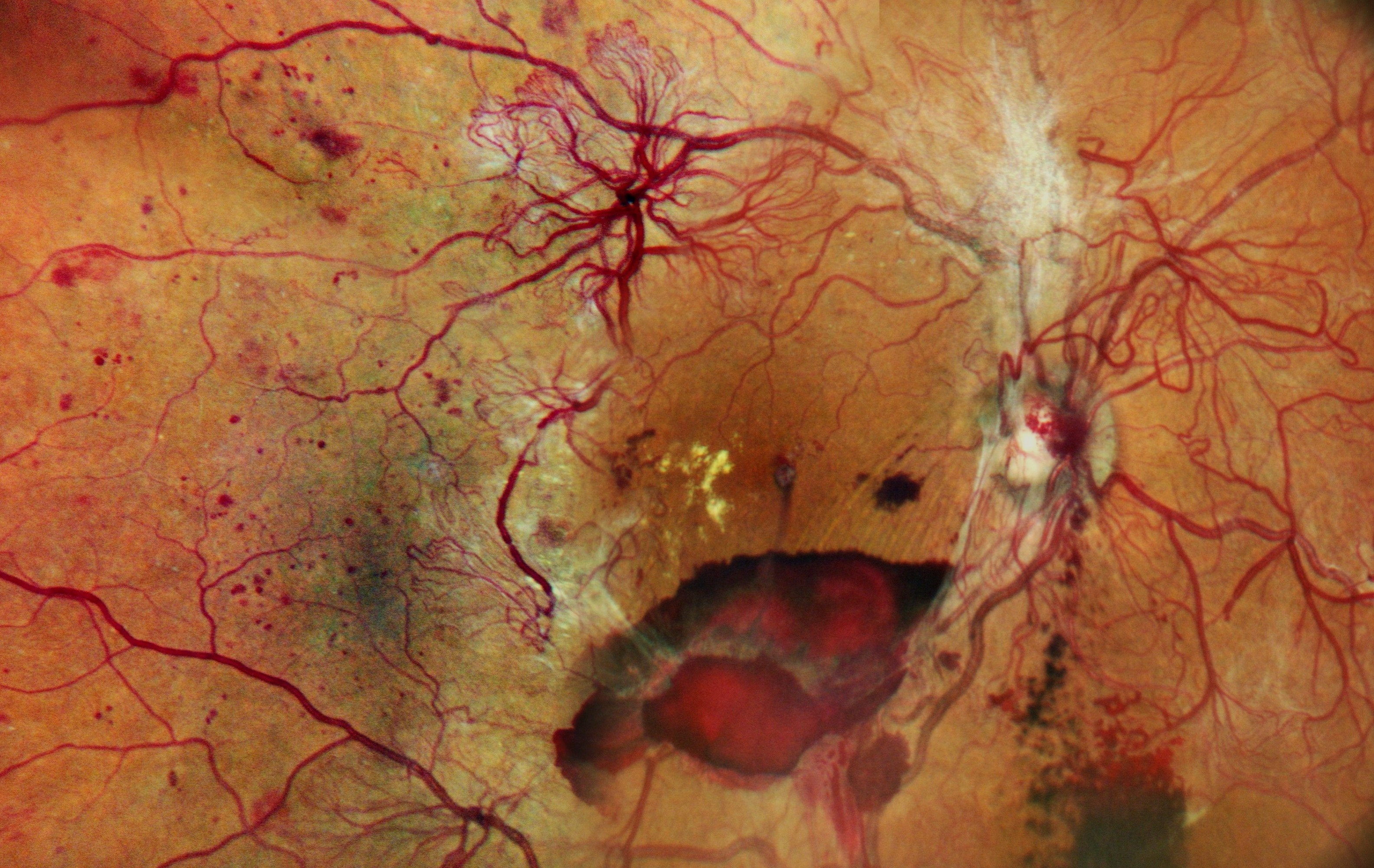
Title: Subhyaloid Rose blooms in Neovascular Garden!
Description: A 50-year-old lady, a known case of uncontrolled diabetes mellitus, presented with diminution of vision in both eyes for 2 months. Visual acuity in both eyes was 6/18. Right eye color fundus photo showed florid NVEs and NVDs, with subhyaloid haemorrhage and minimal VH suggestive of high risk PDR.
Florid Subtype is a hyper-aggressive, fulminant form of PDR. It is characterized by extensive, profuse new vessels spanning several disc diameters in all directions (often described as “coralliform” due to its sea-coral appearance). It progresses over weeks rather than months, carrying a massive threat of sudden blindness.
Dr. Arya A R
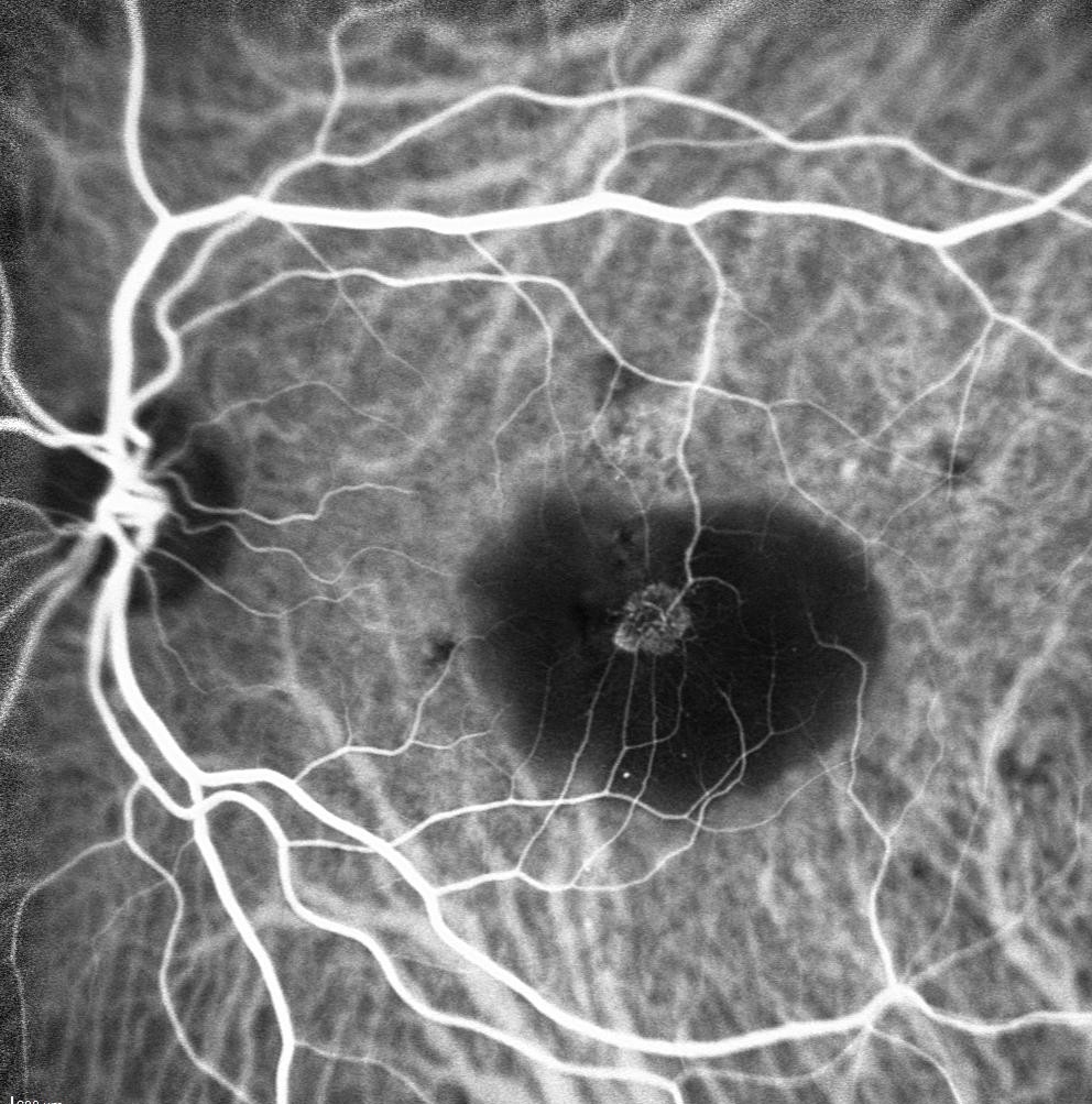
Title: A hairpin loop in the retina’s vascular symphony
Description:76 year old male presented with c/o defective vision in the left eye for the last 3 years. O/E vision was 6/9 (OU). Anterior segment was with in normal limits. Fundus examination of the left eye showed a large PED. OCT showed PED with cystic changes. ICG revealed a hairpin loop with a perfusing retinal arteriole and draining retinal venule suggestive of Retinal angiomatous proliferation. This case highlights the importance of multimodal imaging in such cases.