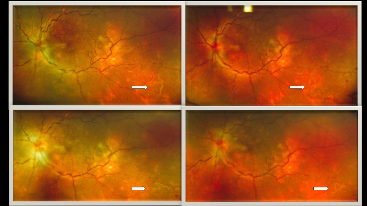
Dr. Farhat Fatima
Title: The chronicle of a wandering worm – DUSN
Description: A 38 year old healthy male presented with blurring of vision in his left eye since 2 weeks, insidious in onset and progressively increased associated with redness since 1 week. Nil systemic. He is a non- vegetarian. His visual acuity in right eye was 6/6 and left eye was CF 1/2m. Anterior segment examination of left eye revealed endothelial dusting, AC cells +1, flare +1, pigments on anterior lens capsule and anterior vitreous phase cells. Fundus examination of left eye showed multiple areas of retinitis involving the posterior pole. In the inferotemporal quadrant, a live motile worm was visualized in subretinal space along with a non- motile worm fragment lying adjacent to it. The worm was highly photosensitive and very motile on IDO examination. OCT of left eye showed gross macular oedema. Diagnosis of Diffuse Unilateral Subacute Neuroretinitis(DUSN) was made. The series of images shows the motility of the worm.
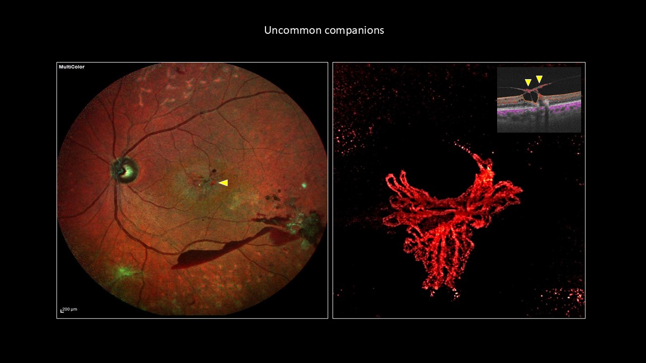
Dr. Shrinivas Shelkikar
Title:Uncommon companions
Description:Multicolor image (left side image) of a 62-year-old male with concurrent proliferative diabetic retinopathy (PDR) and macular telangiectasia (MacTel) displaying intraretinal hyperpigmentation in the perifoveal area and subhyaloid hemorrhage in the left eye. Flow signal within the epiretinal tissue is evident on cross-sectional optical coherence tomography angiography (OCTA) (inset). Color-coded En-face OCTA of the vitreous slab revealed the vascular network (right side image), confirming the presence of foveal epiretinal-neovascularization (ENV). Similar findings were seen even in the right eye. Epiretinal-neovascularisation is a less common finding in eyes with MacTel. The presence of retinal ischemia in PDR may promote the development of foveal ENV in eyes with MacTel.
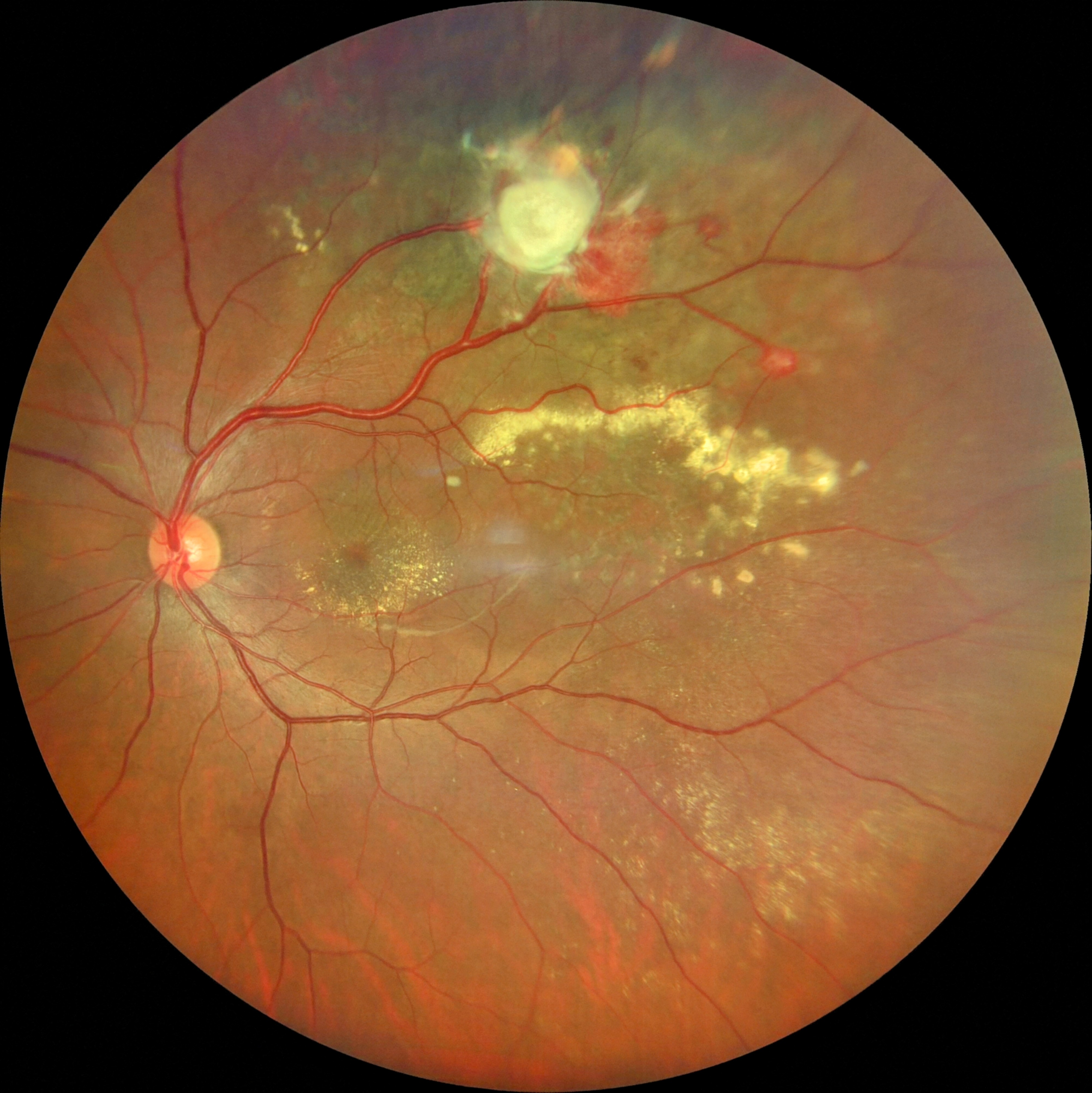
Dr. Keya Chakrabarti
Title: Retinal capillary hemangioblastoma s/p TTT in a patient with VHL
Description: Fundus picture of LE showing normal disc, dilated feeder vessels of the superior arcade. There are multiple hemangioblastomas seen. Exudation seen near the superior arcade that has extended to the macula. The whitish elevation seen superiorly shows a fibrosed hemangioblastoma following trans pupillary thermotherapy.