Dr. Ashutosh Kumar Gupta
Title: Spectacle inside the eye !!
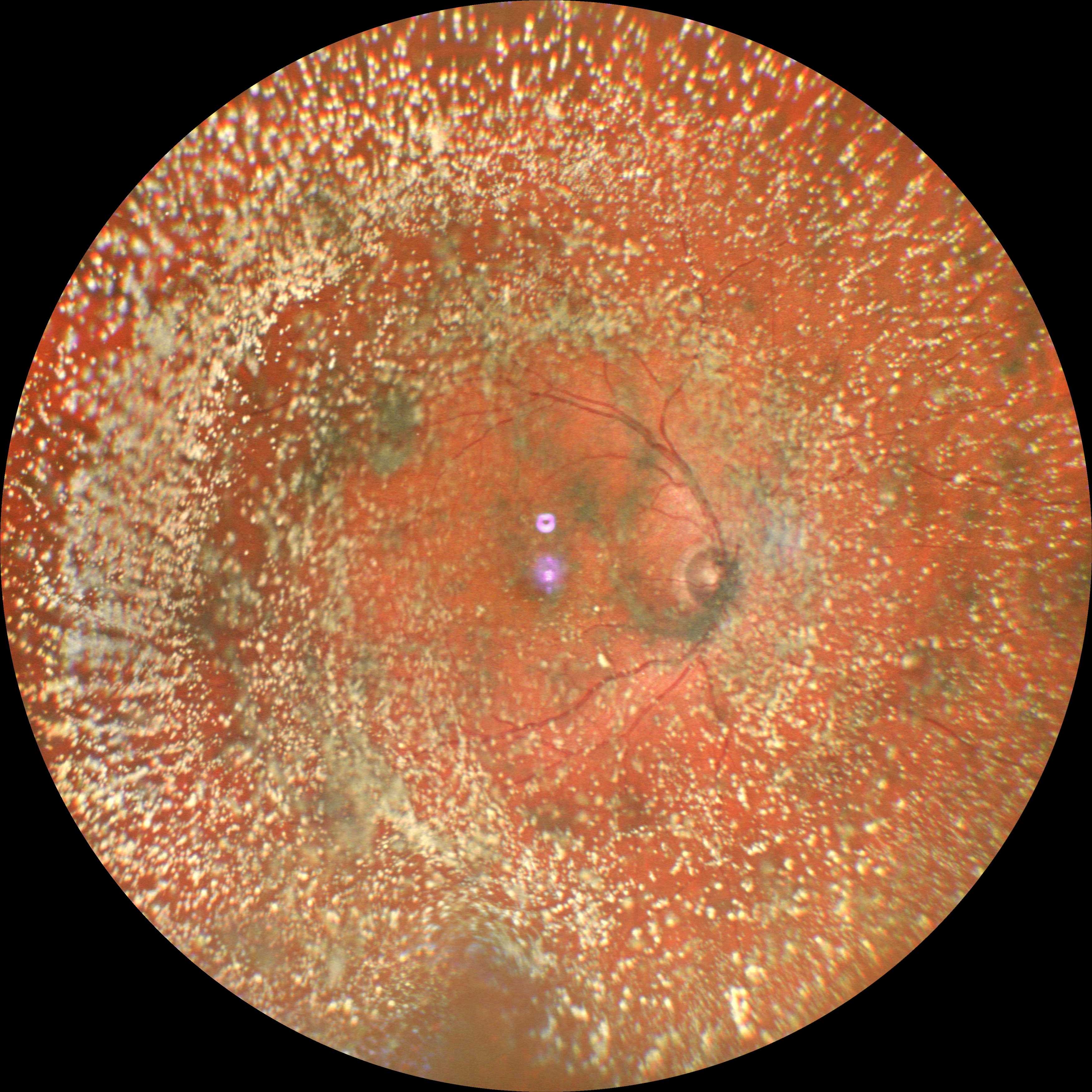
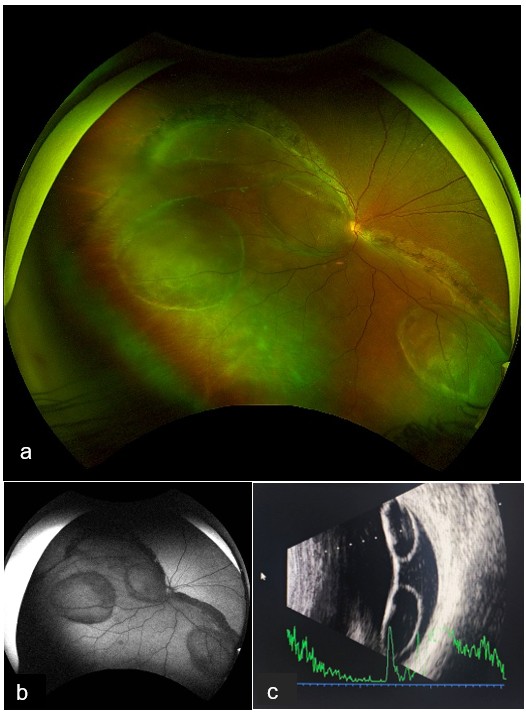
Description: A 40-year-old woman reported a two-year history of progressively declining vision in her right eye, with no prior ocular trauma or corrective spectacle use. Clinical evaluation demonstrated a best-corrected visual acuity of 1/60 in the affected eye and 6/6 in the asymptomatic left eye.
Fundoscopic examination of the right eye revealed an inferior retinal detachment, more pronounced temporally, with macular involvement. Multiple retinal cysts were observed: one at the macula, another inferotemporally, and a third inferonasally.
Figure 1. Ultrawide field pseudo-color image and autofluorescence image of the right eye of the patient with old rhegmatogenous retinal detachment showing multiple intraretinal cysts (a,b). Ultrasound A B-scan showing total retinal detachment with multiple retinal cysts in a chain giving an appearance of a spectacle inside the eye(c).
Dr. Abu Moazzam Parvez
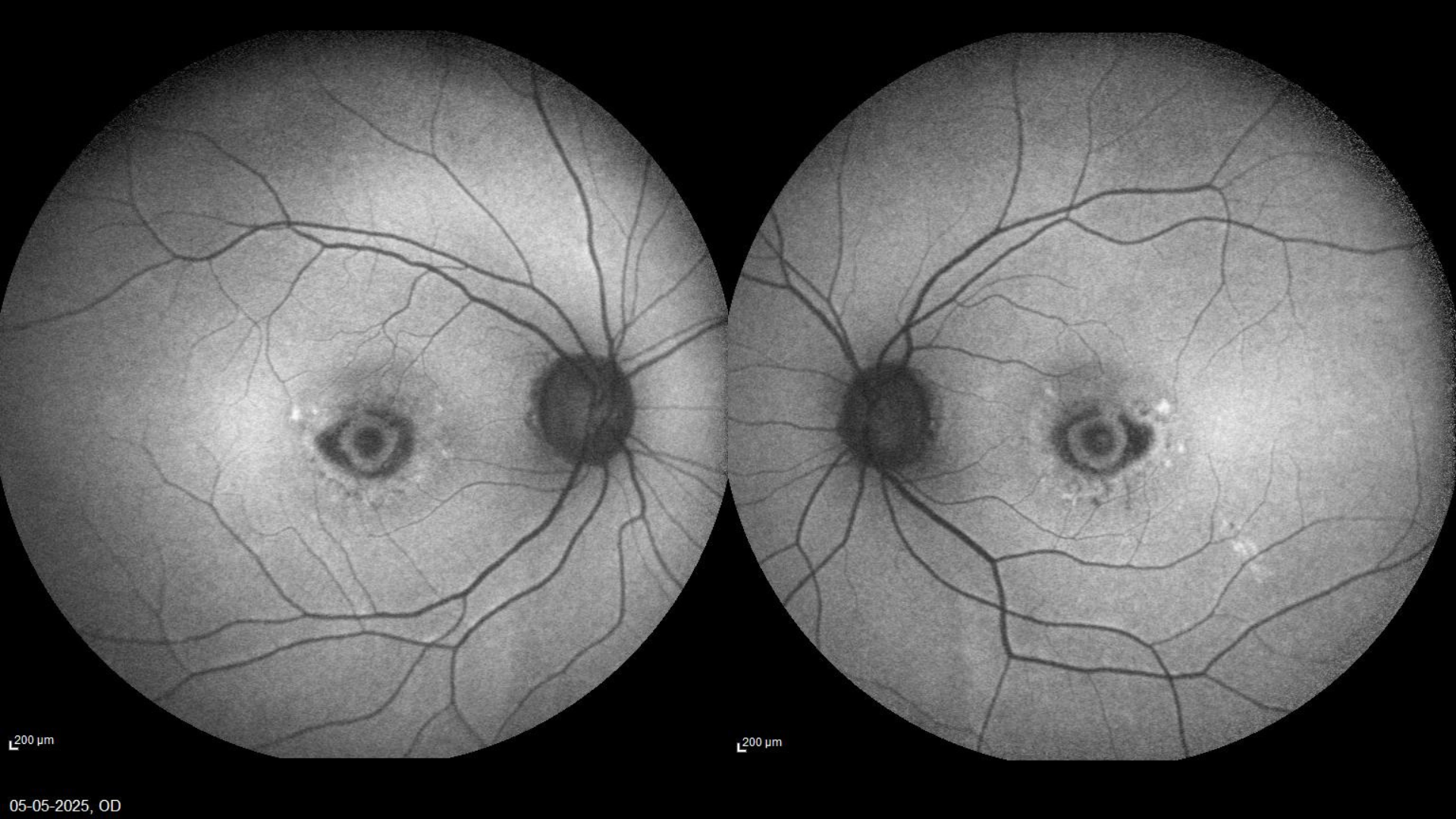
Title: Eye within an Eye : Symmetrical macular dystrophy.
Description:
Bilateral benign concentric annular macular dystrophy (BCAMD).
A 32 y/o woman following up for the past 5 years maintaining a stable vision of 6/6 OU presents with a “bulls eye ” pattern in the macula . In the last follow up patient complains of distortion. On Auto fluoroscense imaging new parafoveal halo was noted. BCAMD is known to develop peripheral vision loss , nyctalopia and retina pimentosa like features in later stages.
Dr. Anjana Mirajkar
Title: Asteroid hyalosis
Description:A widefield image of RE of an 65 year old male with dense asteroid hyalosis.