Dr. Supreme Goel
Title: Built-in Shades: Anatomy Playing Tricks on Oct
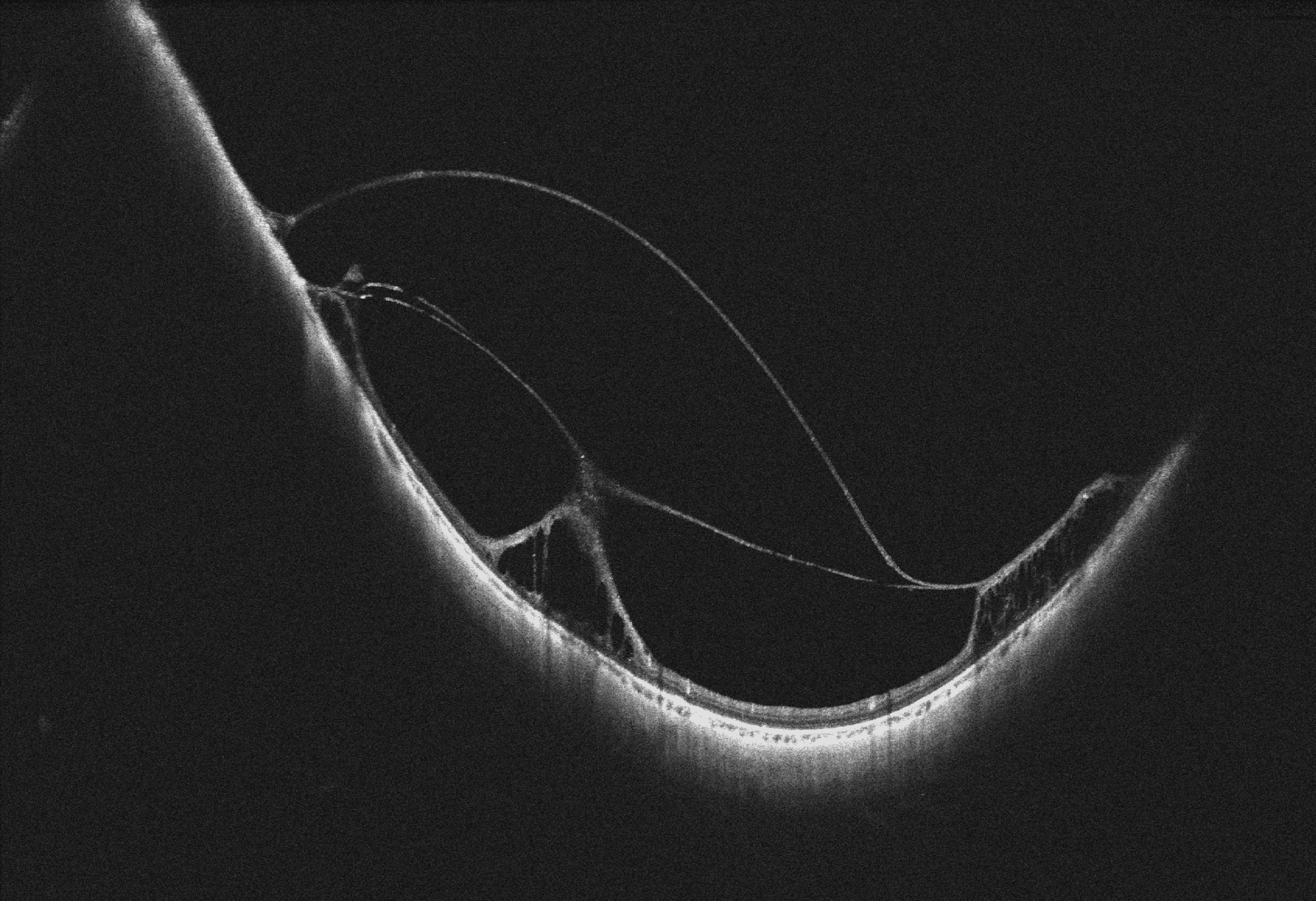
Description:A 29-year-old pathological myopic female, status post belt buckle and vitreoretinal surgery with silicone oil instillation followed by silicone oil removal for subtotal rhegmatogenous retinal detachment, underwent swept-source OCT of the left eye (scan inferior to fovea) revealing incompletely removed vitreous membranes causing tractional schisis. The eye leaves behind traces of its struggle— residual membranes arching into a silhouette that mimics a pair of shades. What appears visually striking is, the biomechanics of traction and schisis at play, yet within this altered contour lies a quiet beauty: anatomy adapting and redrawing itself in the aftermath of trauma.
There was subretinal fluid at macula with vitreomacular traction with tractional schisis and focal tractional detachment. She was taken up for resurgery to relieve the traction.
Dr. Abhishek Gupta
Title: A motile subretinal cysticercus.
Description: “A 35-year-old lady presented with complaints of diminution of vision in the left eye for 1 month. Vision in RE-6/6, LE-6/60. LE Fundus evaluation revealed vitritis. There was a subretinal cysticercus sac superonasal to the optic disc. The scolex was seen protruding outside the sac. Upon strong light stimulation, the scolex retracted inside the sac. The case was taken up for LE pars plana vitrectomy (PPV) and cyst aspiration. 23 G PPV was done. Tricort-assisted posterior vitreous detachment was induced. Complete vitrectomy was done. Then a small retinotomy was made over the cyst with a diathermy. The sac was aspirated out from the subretinal space into the vitreous cavity. It was then aspirated inside the cutter. Fluid air exchange was done. The retinotomy site was lasered. SF6 gas was injected. Postoperatively, the retina was attached.
This is a rare documentation of the motility of the scolex of a cysticercus.”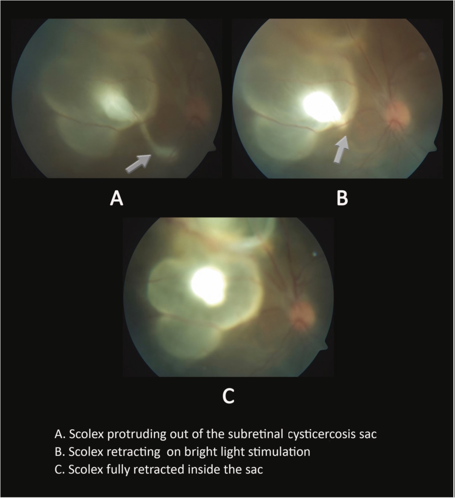
Dr. Manu Sharma
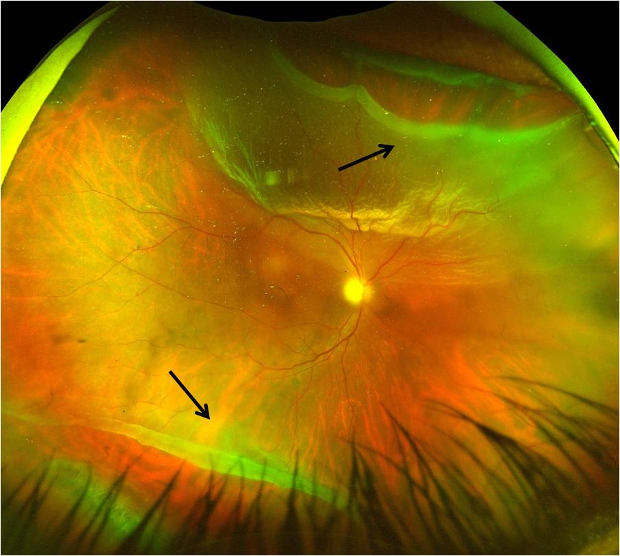
Title: The Visual Corridor between two GRTs
Description: A 59-year-old male presented with a diminution of vision in his right eye. On examination, he had visual acuity of 6/60. Fundus examination revealed 2 giant retinal tears parallel to each other, with the macula attached in between, giving the appearance of a visual corridor between the 2 GRTs. (black arrows in figure)