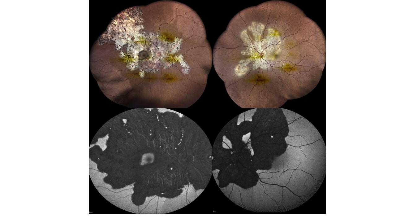
Dr. Supreme Goel
Title: The octopus I beat
Description: Geographic Helicoid Peripapillary Choroidopathy
A swirling octopus of inflammation unfurled across her fundus— pale, relentless tentacles advancing toward fovea, threatening the heart of sight.
Yet vision did not yield: 6/6 in both eyes.
A 34yr/F presented with active disease in her left eye with fovea partly claimed by the advancing helicoid; the right fovea untouched, as shown in fundus images. After steroids, she was maintained on mycophenolate mofetil.
Eighteen months later, autofluorescence images as shown- revealed the creature subdued, its once-furious glow softened into healed serpiginous scars.
The battle left its marks, but not her vision— a quiet triumph of light over the lurking octopus within.
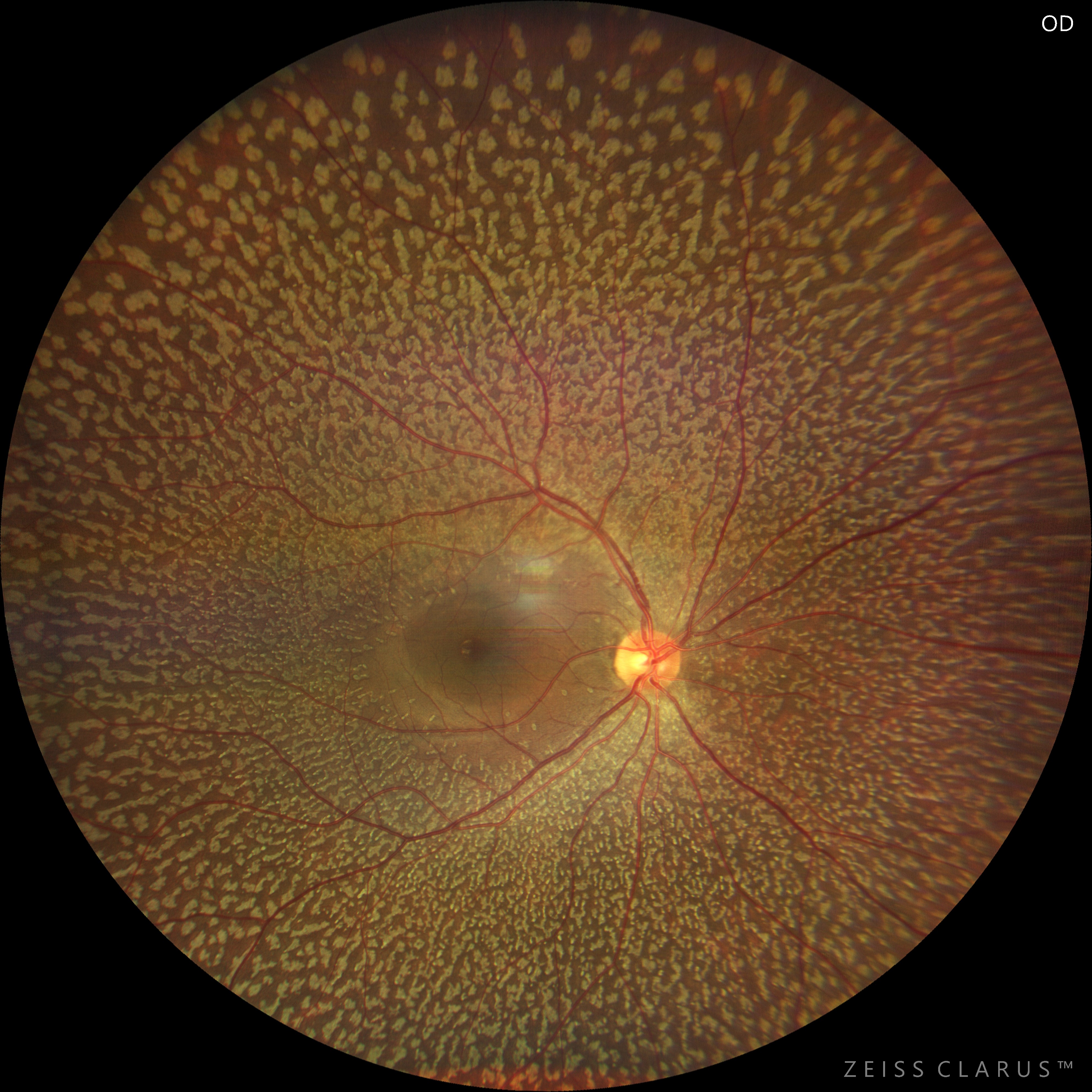
Dr. Akshat B.Kothari
Title: Polka-Dot Retina- The Harmless Pattern
Description: A 32 year female presented to OPD for routine examination. Upon examining the fundus- Multiple flecks were noted throughout the fundus sparing posterior pole. It was bilateral presentation with symmetrical fundus picture in both eyes. OCT and Auto-fluorescence was done for documentation. As patient was asymptomatic, she was advised yearly follow up
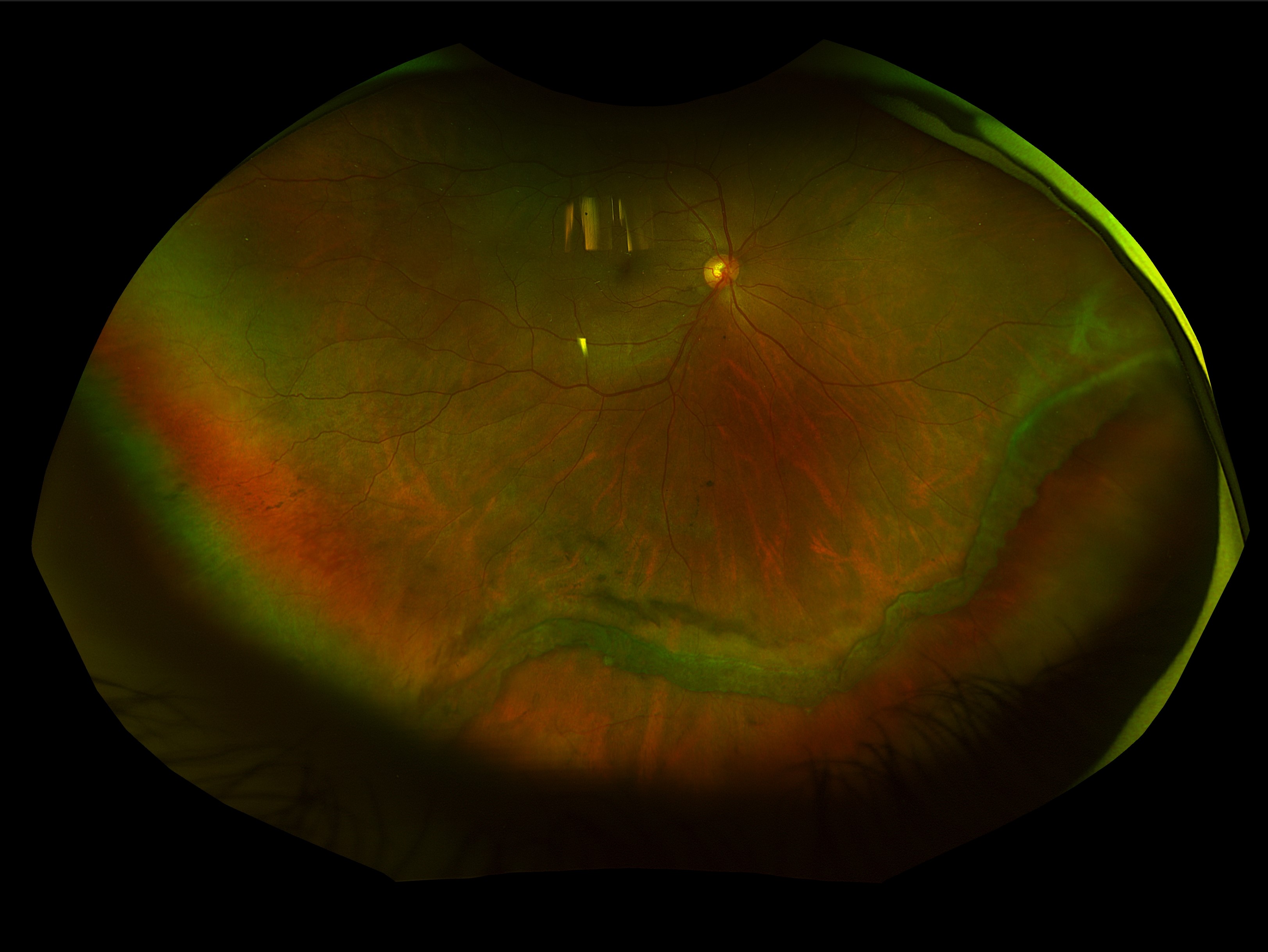
Dr. Catharine Joby
Title: Vitreous base avulsion after closed globe injury
Description: A 32 year old male with history of closed globe injury to right eye 4 months back, presented to us with BCVA- 6/6, N12 in right eye and 6/6, N6 in left eye. Right eye had traumatic mydriasis. Right eye fundus examination showed the vitreous base being detached from the retina from 4-7 o’ clock position, with the retina being attached beneath the avulsion, confirmed by tracing the retinal vessels. It was diagnosed as a case of vitreous base avulsion. This can be a mimicker of retinal dialysis. Patient was managed conservatively with advice for periodic check up.