Dr. Apoorva Chandna
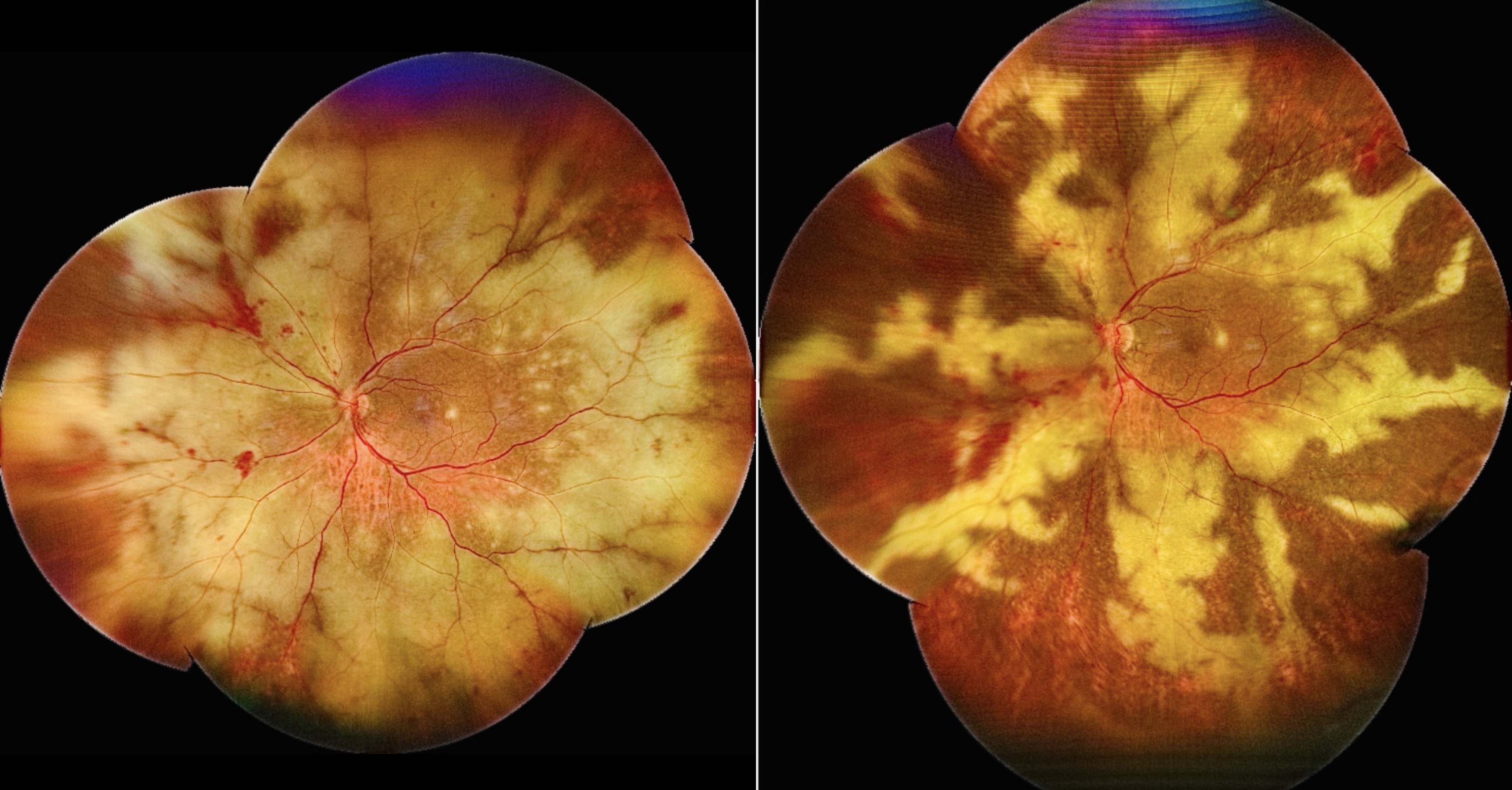
Title: PORN in HIV: A Retina Consumed, Then Contained
Description: Ultra-widefield fundus images depict a case of progressive outer retinal necrosis in an HIV-positive patient, highlighting the stark contrast before and after intravitreal ganciclovir injection.The pre-treatment image shows multifocal, confluent creamy-white outer retinal necrotic patches with rapid circumferential spread, minimal vitritis, and relative vascular sparing—hallmarks of PORN.Post-injection, there is noticeable consolidation of lesions with reduced active borders and early pigmentary changes, suggesting disease control.
Dr. Dasari Maheshkrishnantrivedi
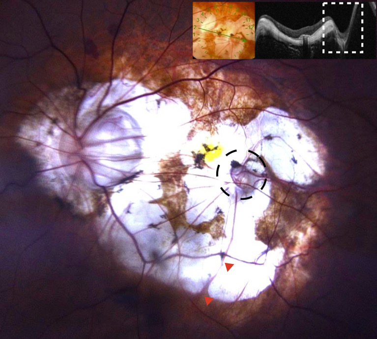
Title: Not a Twin: Pseudoduplication of the Optic Disc Confirmed on OCT
Description: Color fundus photograph of a 56-year-old male with degenerative myopia shows the true optic disc with an additional disc-like structure at the macula (dotted black circle). Retinal vessels are seen emerging from the true disc. The vessels emerging from the pseudo disc are choroidal vessels as seen by its presence at a deeper level beyond the area of chorioretinal atrophy (red arrowhead). Optical coherence tomography through the pseudo disc demonstrates excavation of sclera and preserved attenuated retinal tissue at the base. These findings indicate that the macular pit at the point of entry of short ciliary artery is appearing as pseudodisc.
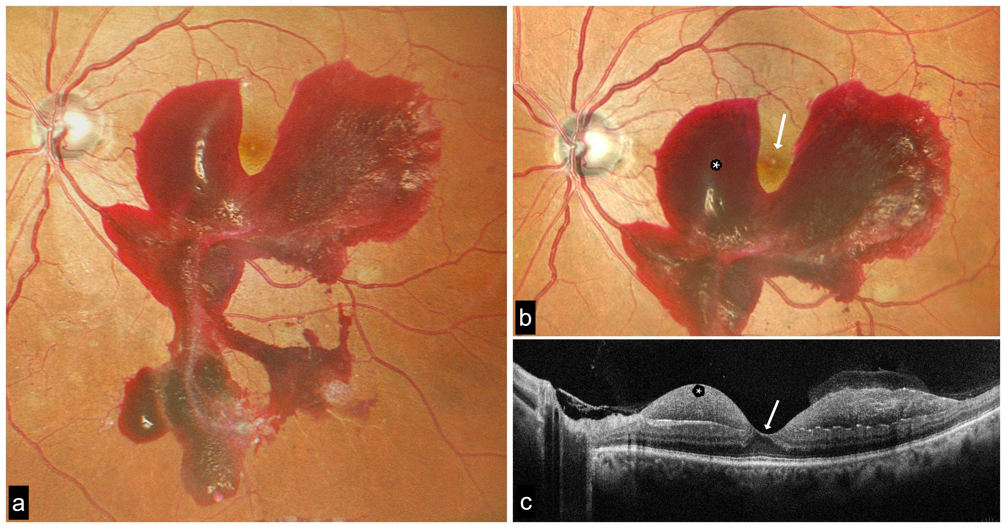
Dr. Shraddha Raj Shrivastava
Title: Foveal Refuge amidst the Subhyaloid Flood
Description: A middle-aged man with poorly controlled type 2 diabetes mellitus, having left eye visual acuity of 20/20, presented with a large premacular subhyaloid haemorrhage extending beyond the inferior arcade, with complete foveal sparing despite extensive accumulation of blood. OCT demonstrated a perifoveal dome-shaped subhyaloid haemorrhagic cavity with preservation of the foveal contour and intact outer retinal layers.
The distinct distribution pattern is consistent with the known sequence of posterior vitreous detachment, wherein vitreous separation begins in the perifoveal region before involving the fovea, leaving persistent vitreo-foveal adhesion that may restrict haemorrhage from tracking into the foveal center.
The striking anatomical configuration of foveal sparing despite extensive premacular hemorrhage highlights the in-vivo mechanical influence of staged posterior vitreous detachment and vitreoretinal interface dynamics in determining the spread of subhyaloid hemorrhage.