QUICK LINKS
Winner of April, May & June 2022

Dr. Akansha Sharma
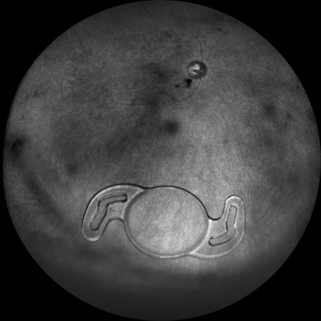
Description: INFRA-RED FUNDUS IMAGE OF A 47 YEAR OLD MALE WITH INTRA-OCULAR LENS IN THE VITREOUS CAVITY OF THE RIGHT EYE.
Dr. Rushik Patel
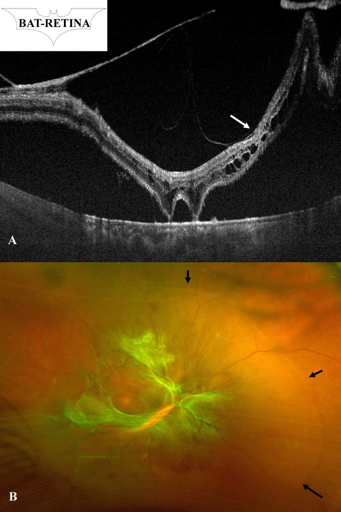
Description: BAT-RETINA: The swept-source optical coherence tomography of 45 year old diabetic female having treatment-naiive proliferative diabetic retinopathy with combined retinal detachment (black arrows) (B) in her right eye showed tractional retinal detachment involving macula (white arrow) except 2 focal attachments with underlying retinal pigment epithelium (A). This appearance resembles BAT-RETINA”.
Dr. Harshit Vaidya
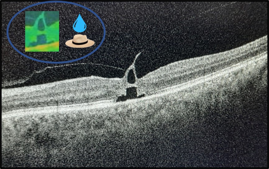
Title : Tear drop on a hat sign: Foveal pseudocyst atop a splayed outer macular hole in a case of Vitreo-Macular Traction
Description: Optical Coherence Tomography (SD-OCT) image of a 59-year-old female complaining of diminution of vision in the left eye for 8 months (best corrected vision CF 1 metre). Left eye revealed vitreo-macular traction causing a foveal pseudocyst (tear drop) separated by a bridging retinal tissue and placed atop an outer macular hole with a splayed base (hat) and disrupted ellipsoid zone. The detaching posterior hyaloid exerts vertical traction over the temporal aspect and horizontal traction over the fovea nasally. The pull of the vertical vector appears to be greater than horizontal as evidenced by the vertically elongated pseudocyst and broad base of the outer hole.

Dr. Jiz Santhosh
Description: Macular Fireworks

Dr. Roshni Mohan
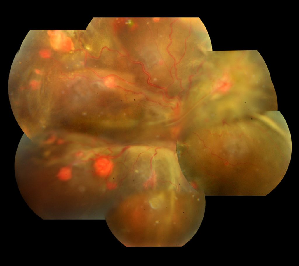
Title : Multiple Berries in the Eye
Description: A 21 year old female, monocular patient reported to our OPD with complaints of defective vision in her only seeing eye. At presentation , her BCVA OD was 6/18, OS NLP. Anterior segment examination was unremarkable OD. Fundus evaluation OD had clear media with hyperemic disc , inferior exudative RD and multiple dilated reddish elevated lesions in the mid periphery having a feeder arteriole and draining venule. Multiple small to medium sized reddish elevated lesions were seen throughout the fundus. Inferior and nasal exudative RD was noted.A diagnosis of multiple retinal capillary hemangioma(RCH) , probable Von Hippel Lindau’s Disease(VHL),complicated by exudative RD in a monoocular patient was made.
Dr. Bhavani S
Title : Full thickness macular hole secondary to Best vitelliform dystrophy
Description: The fundus photograph of the right eye of a male patient aged 18 years, who presented with Full thickness macular hole secondary to Best vitelliform dystrophy


Dr. Mahak Bhandari
Title : Multimodal Imaging of Bilateral Symmetrical Basal Laminar Drusen
Description: A 62-year-old male patient presented to us with diminution of vision in right eye for 6 years. Multicolor picture showed us well demarcated large drusen present bilaterally and symmetrically. OCT revealed multiple pigment epithelial detachments (PED) with steep sides and hypo reflective homogenous content bilaterally. The drusen coalesced subfoveally in the right eye along with RPE and photoreceptor disintegration justifying the poor vision. FFA showed typical starry sky appearance starting from the arterio venous phase and ICG showed hyper cyanescens surrounded by hypo cyanescens due to RPE atrophy at the peak of the PEDs.
Multimodal imaging of the patient confirms an atypical presentation of basal laminar drusen in terms of larger size which should not be confused with serous PEDs.

Dr. jiz santhosh
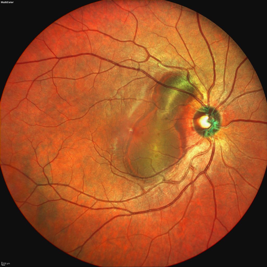
Title : Hemorrhagic Pigment Epithelial Detachment
Description: A multicolour image of the right eye showing a hemorrhagic PED secondary to a peripapillary polypoidal choroidal vasculopathy.
Dr. Kushal Delhiwala
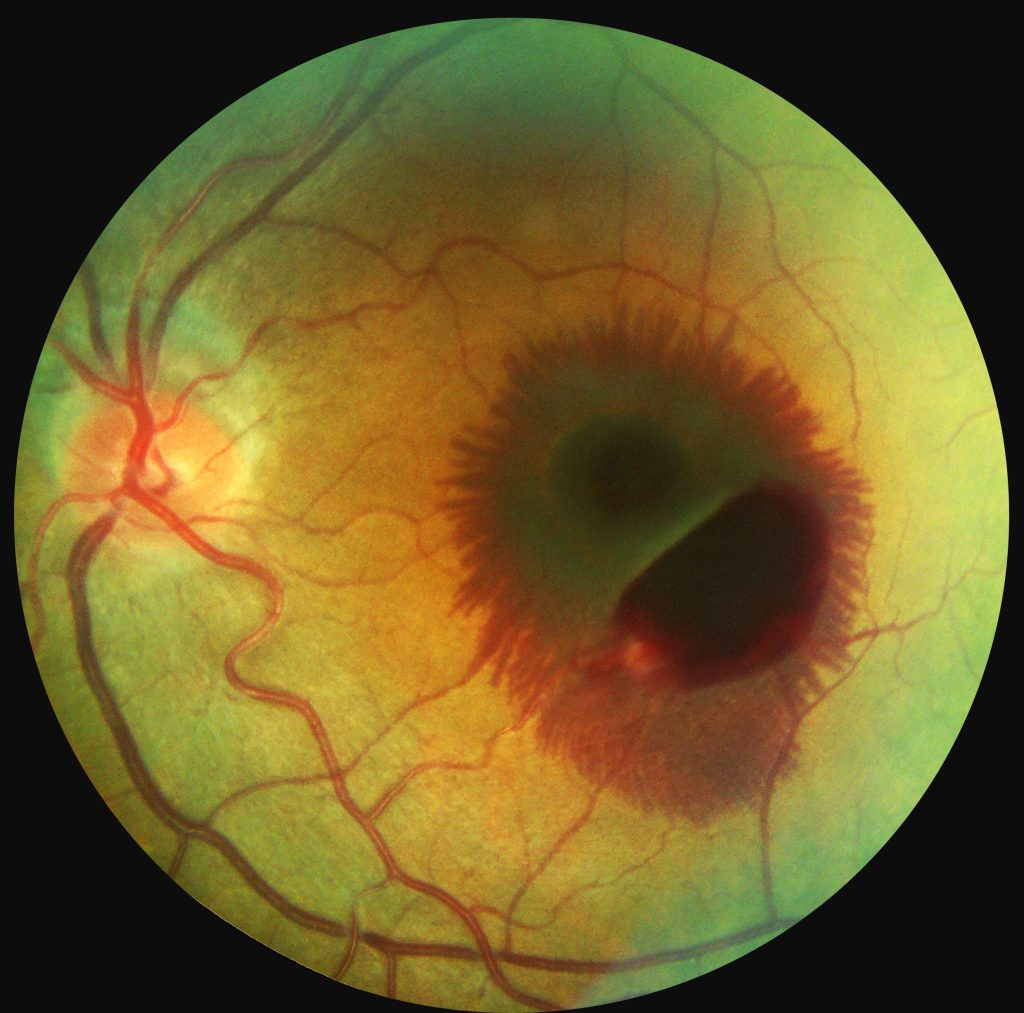
Title : Preretinal hemorrhage with radial hemorrhage in Henle’s fibre layer
Description: A 30-year-old phakic male presented with left eye central blurring of vision for 4 days. Fundus photograph of posterior pole showed preretinal hemorrhage with radial Henle’s fibre layer (HFL) hemorrhage at macula. On evaluation, patient was diagnosed to have anemia and referred to physician for further management. HFL hemorrhage can occur secondary to abnormal retinal venous pressure affecting the deep capillary plexus or from choroidal vascular pathologies. Radial appearance of HFL hemorrhage results from peculiar anatomical arrangement of the photoreceptor axons and thus hemorrhage gets accumulated along the direction of fibres.
