QUICK LINKS
Retinal vascular occlusion
What is Retinal vascular occlusion?
The blood supply of the retina consists of arteries and veins which carry blood to and from the retina back to the heart. This serves as the main supply of nutrition to the retina. Any blockage in these vessels is termed as vascular occlusion. It can be termed as retinal vein occlusion and retinal arterial occlusion.
Retinal vein occlusion (RVO) is more common than arterial occlusion and can be either central or branch retinal vein occlusion. Occlusion can be either be due to clot in the veins or due to high blood pressure. Vision can be blurred because of 3 reasons:
– Macular edema – buildup of fluid or leakage in the central part of the retina
– Haemorrhages – blood in the retina or blood in the vitreous cavity
– Ischemia – lack of blood supply to the retina/macula due to blockage


What are the signs and symptoms and risk factors for RVO?
Signs and symptoms can vary from sudden painless loss of vision to blurring of vision. There can be dark spots or floaters in the visual field. Risk factors are uncontrolled hypertension and diabetes, smoking, obesity, raised cholesterol and advanced glaucoma. In young adults, inflammatory conditions and clotting disorders are more commonly associated with retinal venous occlusions than in older population.

I have been diagnosed with central retinal vein occlusion and the retina specialist has advised OCT and Fluorescein angiography as further tests. Are they necessary?
Optical Coherence Tomography (OCT) and Fundus Fluorescein Angiography (FFA) are retinal investigations necessary to further evaluate the condition in detail.
FFA gives us more information about the blood supply of the retina – whether the retina is well perfused or the circulation is compromised. Also, FFA is useful to detect the presence of neovascularization. OCT is required to detect macular edema and is useful to assessing the efficacy of treatment. There is a newer investigative modality called as OCTA (OCT angiography) which is used to see the retinal circulation without the use of dye and is especially important in patients who are allergic to the dye or in whom the FFA is contraindicated.

The doctor has advised some injections in the eye. Is there any other treatment that can work? How many injections will I need?
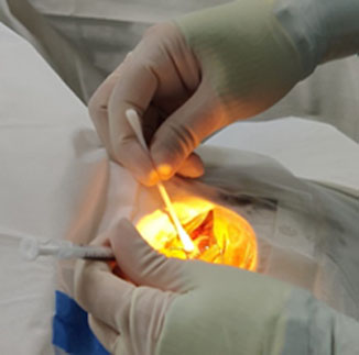
In order to treat the macular edema, the retina specialist advises intravitreal injections which are aimed to reduce swelling and improve the vision. Depending on the extent of damage to the circulation and associated systemic factors, the number of injections can vary. However, since the response to treatment can vary from patient to patient, it is not possible to predict the number of injections required and it is necessary to be ready for multiple repeated injections.

I was diagnosed with CRVO 6 months back and now my eye pressure is increasing. Why is the retina specialist advising laser treatment in addition to injections?
CRVO can predispose a patient to develop abnormal new blood vessels in the retina and in the angle of the eye which can cause eye pressure to increase. This condition is called as neovascular glaucoma and is seen in ischemic CRVO. The laser treatment is done in 3-4 sittings to burn the retina which has lost its blood supply to reduce the new blood vessels and prevent bleeding in the eye. In very advanced cases, the patient may need to undergo glaucoma surgery to bring the pressure under control if it is not controlled with medications.

How much will my vision improve after the laser and injections? Will surgery help?
Visual improvement will depend on the health of the retina and amount of swelling in the macular area. Vitrectomy surgery is required when there is bleeding in the vitreous to clear the blood and improve the visual acuity.
In addition to all these treatment modalities, lifestyle modification such cessation of smoking and control of systemic factors like BP, DM and cholesterol play a very important role in the prognosis and need for continued intervention.

