Dr. Shikhara Vijayan
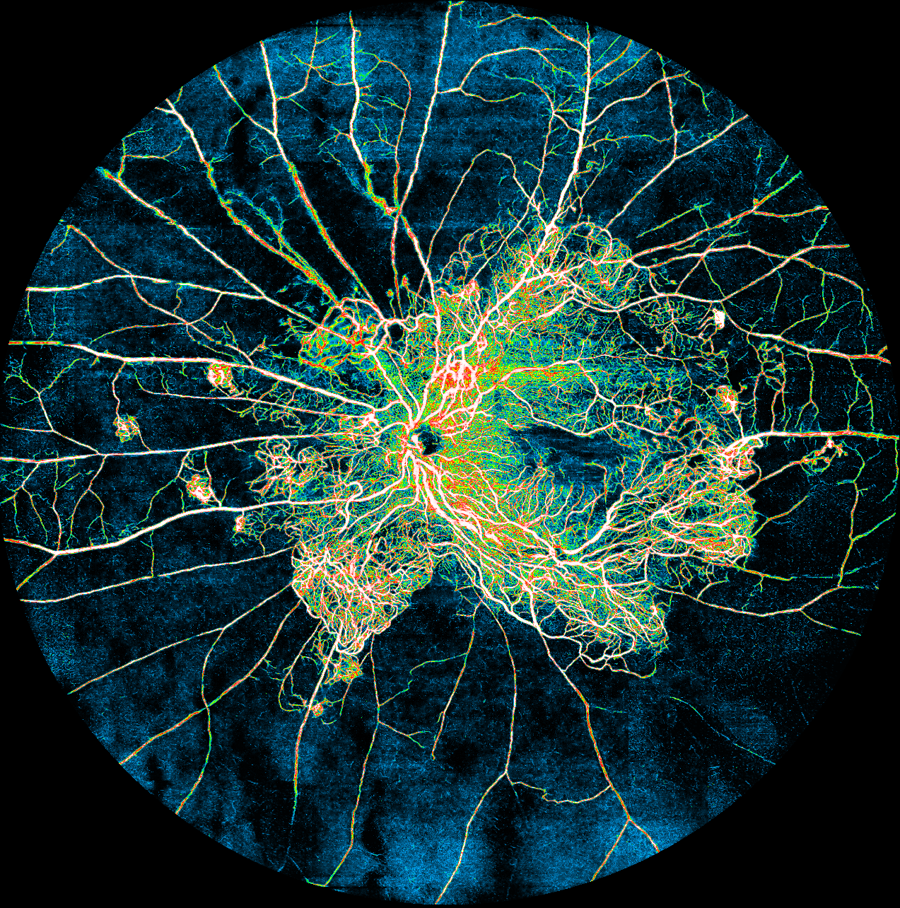
Title: The Withering Vascular Tree
Description: left eye OCT Angiography image of a 32 year old man with Both Eyes Advanced Diabetic Eye Disease with extensive sclerosed vessels.
Dr. Tanya Verma
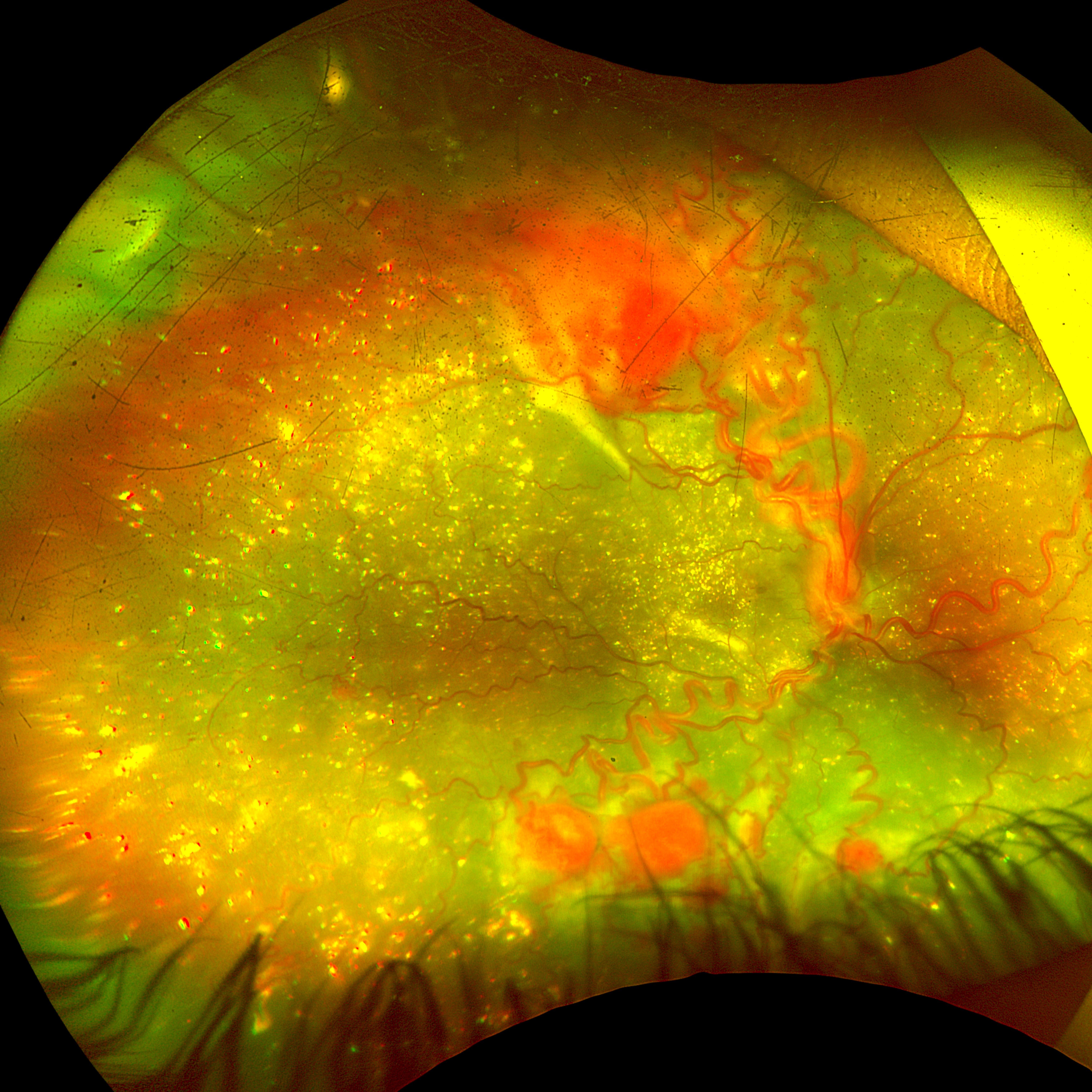
Title: Constellation of Angiomas
Description: This ultrawide-field pseudocolour fundus photograph reveals multiple retinal capillary haemangioblastomas scattered across the posterior pole and periphery, appearing as vivid orange-red nodular lesions with prominent feeding arterioles and draining venules. Set against this vascular backdrop, innumerable lipid crystal deposits glisten throughout the vitreous cavity refracting light as a constellation of golden specks.
The combination of florid angiomatous proliferation and vitreous lipid crystallisation creates a rarely captured funduscopic tableau, likely in the setting of Von Hippel-Lindau disease. This image stands as a testament to the hidden beauty within pathology.
Dr. Adithya Shankar
Title: A Rare Dual Vascular Catastrophe
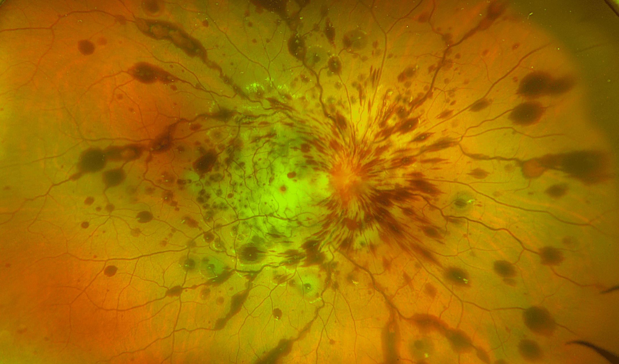
Description: A 15 year old girl presented with loss of vision in RE following trauma with a wooden stick. O/E: She perceived no perception of light in RE and RAPD was present.
Fundus optos image showed diffuse whitening of retina with absent central cherry red spot suggesting Ophthalmic Artery Occlusion (OAO) along with extensive flame and blot haemorrhages throughout the fundus explaining possible venous outflow obstruction due to severe disc edema.