Dr. Vipin rana
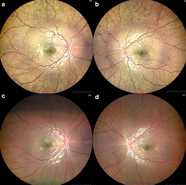
Title: The Vanishing Golden Fundus: Mizuo–Nakamura Phenomenon in GRK1-Associated Oguchi Disease
Description: A 6-year-old boy was referred with a one-year history of nonprogressive night blindness. Parents noted difficulty navigating in dim light. BCVA was 20/20 OU. The anterior segment examination was normal. Fundus examination showed a diffuse metallic golden sheen bilaterally (Figure 1a,b). Optic discs and vessels were normal. After prolonged dark adaptation of 03 hours, the abnormal sheen resolved completely, revealing a normal fundus, consistent with the Mizuo Nakamura phenomenon (Figure 1c, d). Full-field ERG showed markedly reduced scotopic rod responses with relatively preserved photopic cone responses, suggestive of congenital stationary night blindness. Genetic testing revealed compound heterozygous pathogenic variants in the GRK1 gene involving exons 3 and 7. Segregation analysis confirmed autosomal recessive inheritance. Parents were counselled regarding the benign, nonprogressive course and a 25 percent recurrence risk
Dr. Gaurang Sehgal
Title: Fireball of myelin.
Description: A 65-year-old monocular patient presented with sudden, painless decrease in vision in the OD. The OS had previously lost vision due to an untreated long-standing RD. At presentation, best-corrected visual acuity in OD was HM+ Color fundus revealed features of CRAO, including diffuse retinal whitening and edema involving the posterior pole, indicative of inner retinal ischemia. Notably, a patent cilioretinal artery was seen arising from the peripapillary region, supplying the macular area. These findings were well corroborated on spectral-domain OCT, which demonstrated preserved retinal architecture in the macular region perfused by the cilioretinal artery, while the remaining retina showed inner retinal hyperreflectivity and thickening consistent with ischemic changes.
An urgent anterior chamber paracentesis with gonioscopic ocular massage was performed. Owing to cilioretinal artery sparing, the patient showed visual recovery, with visual acuity improving to 6/36 at 14-day follow-up. 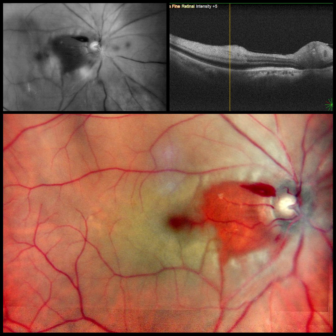
Dr. Apoorva chandna
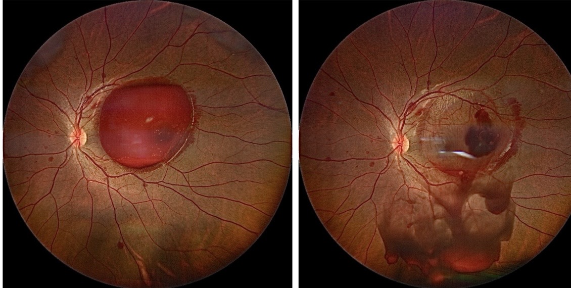
Title: Fall of the crimsom dome
Description: Image showing premacular subhyaloid hemorrhage in Valsalva retinopathy before and immediately after Nd:YAG laser hyaloidotomy — converting a vision impairing premacular collection into a resolving vitreous hemorrhage.