Dr. Greeshma M G
Title: Fundus caldera.
Description: Wide field fundus image showing post traumatic combined retinal and choroidal detachment with the posterior pole forming the central floor of a volcanic caldera like appearance.

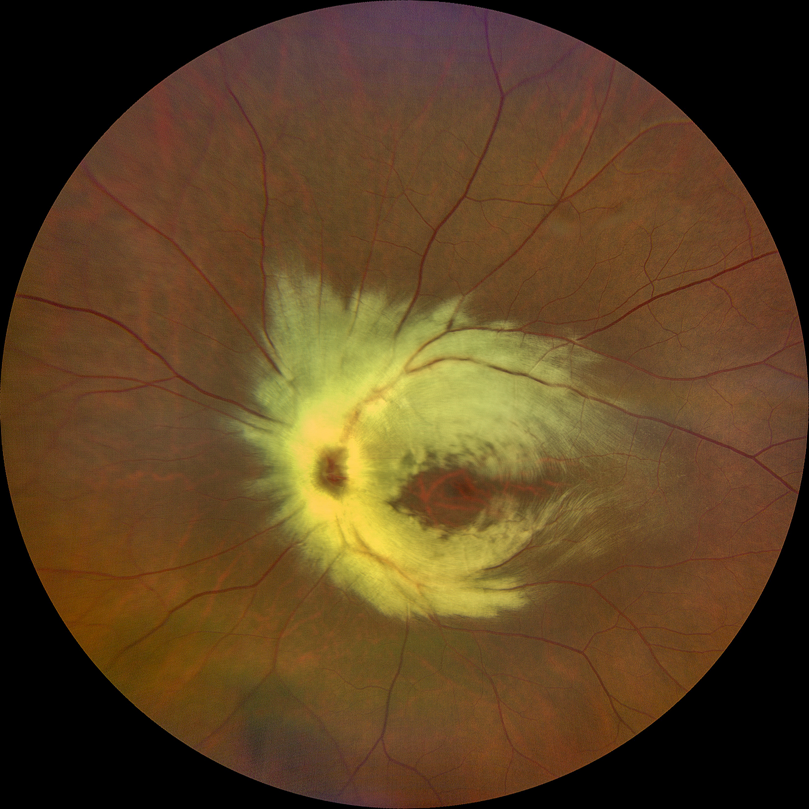
Dr. Mihir
Title: Fireball of myelin.
Description: A 35-year-old male presented with defective vision in the left eye since childhood. Uncorrected visual acuity in the left eye was 6/60 which improved to 6/24 with a correction of -3.0DS. Fundus examination of the left eye showed a tessellated background, and extensive myelination of retinal nerve fiber layer around the disc and arcades. Optical coherence tomography showed a myopic contour with normal retinal layers at the fovea. The right eye of this patient was emmetropic with an unremarkable fundus. He was diagnosed with a triad of unilateral myopia, medullated nerve fibers and amblyopia in the left eye.
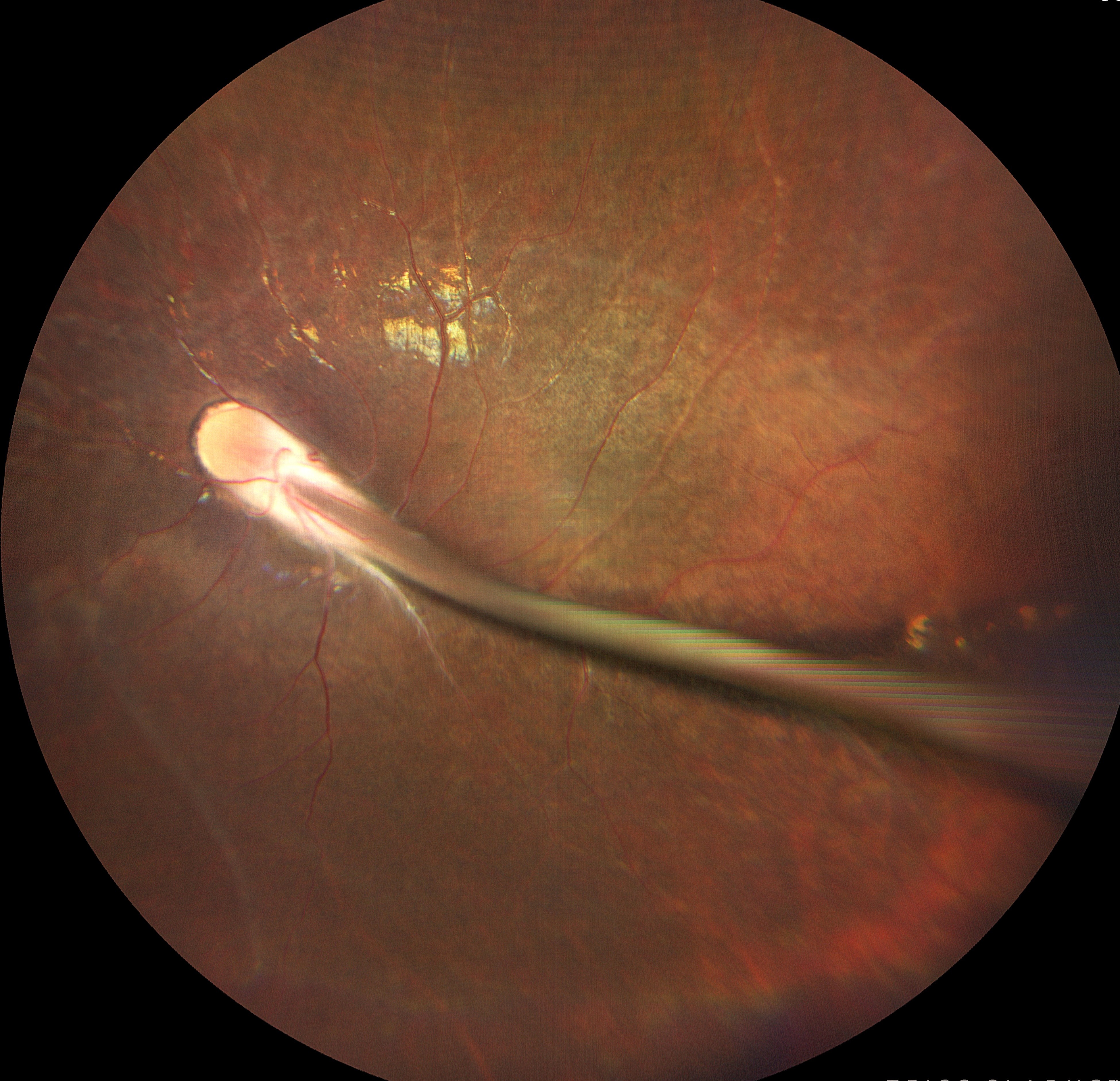
Dr. Giriraj Vibhute
Title: Frozen in time- The portrait of persistent fetal vasculature.
Description: Picture showing fundus of a 19 year old amblyopic girl. Persistent fetal vasculature/ Persistent hyperplastic primary vitreous is due to failure of regression of fetal vessels in the eye. It is the second most common cause of infantile leukocoria. These children are prone to develop amblyopia, cataract, glaucoma, retinal detachment.