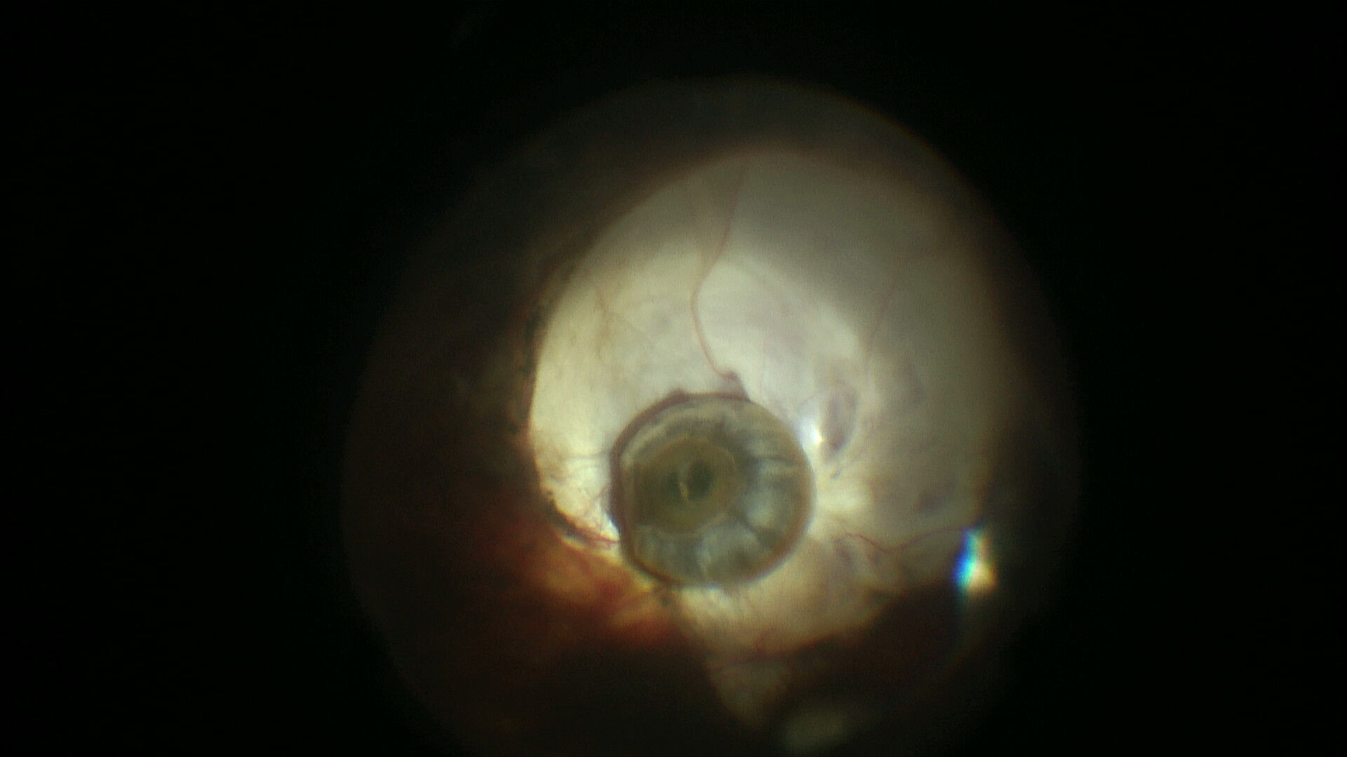
Dr. Manu Sharma
Title: Snail shell appearance of Cataracta Nigra
Description:
A 50-year-old Asian female with iridofundal coloboma and a history of left eye post-traumatic vitreous hemorrhage. Upon examination, her visual acuity was limited to light perception, with normal intraocular pressure. Posterior segment details were obscured due to a grey vitreous hemorrhage. B-scan ultrasonography revealed vitreous hemorrhage with posterior subluxation of the nucleus.
The intraoperative image, post-clearance of the vitreous hemorrhage, shows a black, round structure resembling a snail shell, which was identified as a nucleus (cataracta nigra) positioned on the coloboma surface. This was successfully removed using a phacofragmentome, and pars plana vitrectomy was completed. Post-surgery, the patient’s visual acuity improved to 6/60.
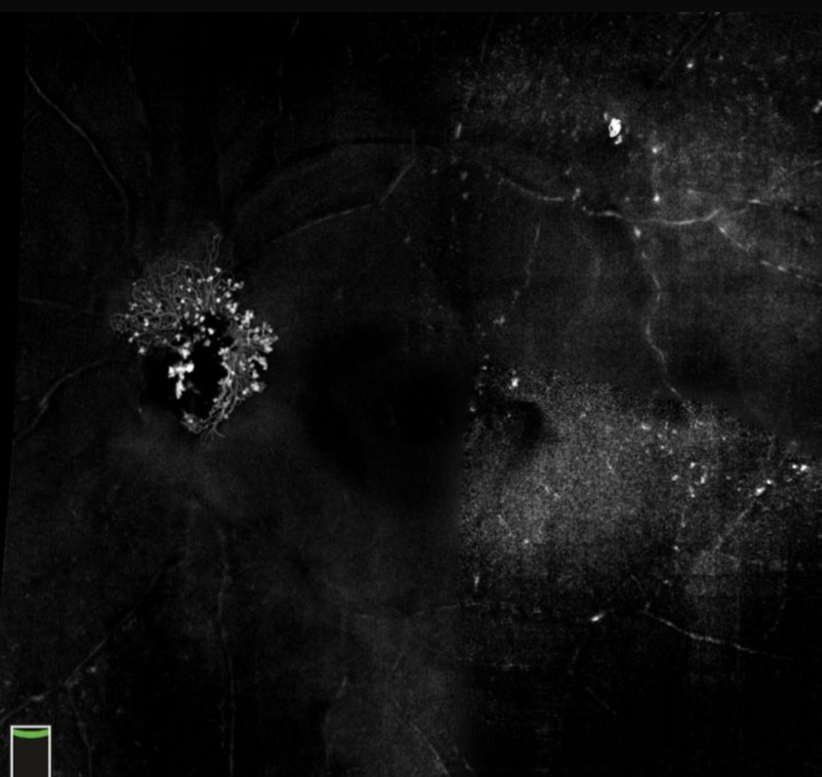
Dr. Aaditi Anilkumar
Title: Nebula on the Nerve
Description:Its a solix 12 mm OCTA scan – Enface mode that shows the neovascularistation of disc in a patient with multiple brvo.
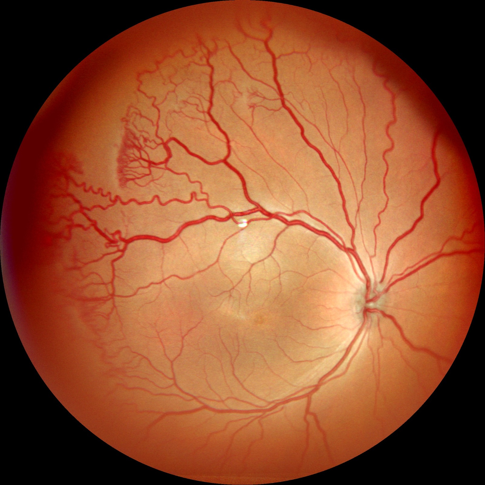
Dr. Deepanshu Agrawal
Title: Widefield Glimpse of A-ROP
Description: An image of the right eye of a premature newborn, born at 32weeks and weight of 1550gms, presented for ROP screening on 21st day at SNCU. On dilated evaluation baby was noted to have dilated and tortuous vessels along with flat neovascular network at the junction between the posterior vascularized and non-vascularized retina. The baby was diagnosed to have Zone 1 Aggressive ROP in both the eyes and was treated with immediate bilateral intravitreal Anti-VEGF. Image Captured: Nidek, Mirante!