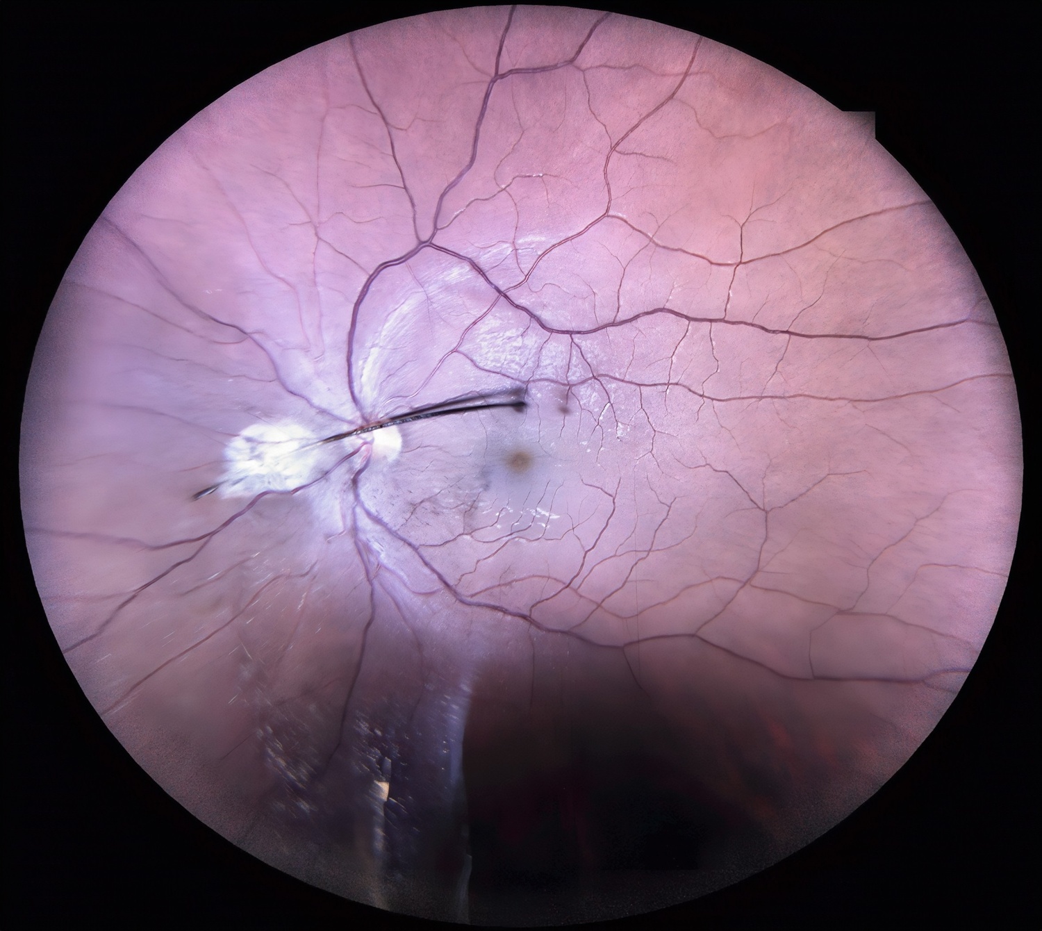
Dr. Anugraha B
Title: Eyelash Migration Into the Eye – When beauty meets the unexpected!
Description: This case highlights an intriguing occurrence of eyelash migration into the eye due to trauma, as evidenced by fundus photography.
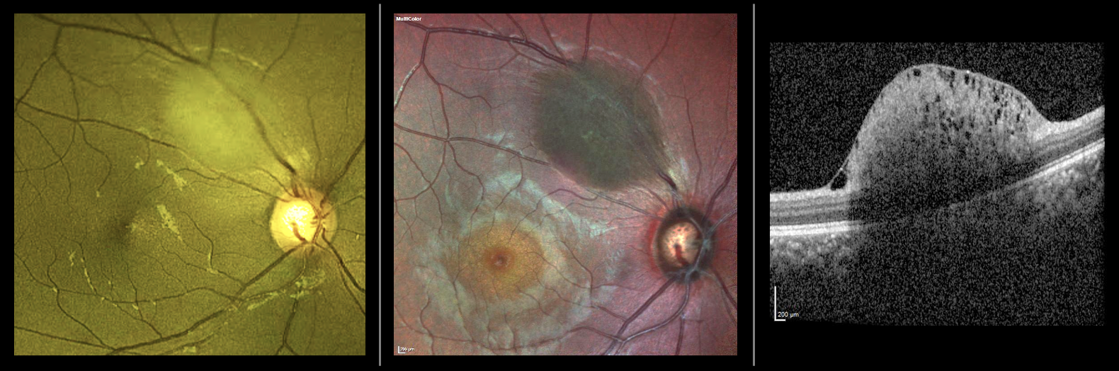
Dr. Apoorva Guruprasad Ayachit
Title: Not just any hypopigmentation in the retina!
Description:
A 27-year-old woman presented with headache. Vision was 6/6 N6 in both eyes. Incidentally a yellowish- white ovoid lesion was seen along the superotemporal arcade in the right eye. An OCT through the lesion showed an elevated lesion with small cystic spaces (moth- eaten appearance), arising from the nerve fibre layer with mild back shadowing. These features were consistent with a type 1 non-calcified retinal astrocytic hamartoma (RAH). Neuroimaging was advised and tuberous sclerosis complex (TSC) was ruled out by a physician and radiologist. RAH when associated with TSC are frequently bilateral, multifocal and may show calcification. Sporadic, solitary type 1 RAH, like in our case, are relatively flat, smooth, semitransparent, yellowish-white lesions without calcification. They are incidentally picked up on routine examination and are unlikely to progress.
Dr. Tejaswita Verma
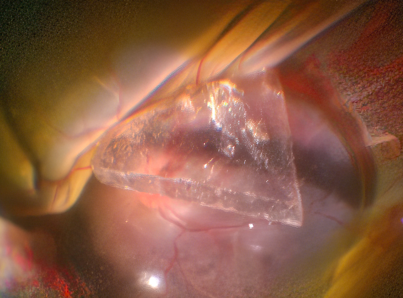
Title: Intraocular glass piece
Description:Intraoperative still of an intraocular foreign body(glass piece) with retinal detachment