QUICK LINKS
Winner of July 2021

Dr.GAGANJEET SINGH GUJRAL.
Abstract
Image shows modified immersion B scan with the help of water balloon depicting posterior displacement of the lens with intact anterior and posterior capsules. This technique helps in delineating details of anterior structures like lens and iris which a conventional B scan would fail to do. This modified technique is also useful in cases of trauma where Immersion B scan/ Ultrasound biomicroscopy with a cup is not recommended.

DR.KSHITIJ S TAMBOLI
Vitreoretina fellow, R.I.K , Bangalore
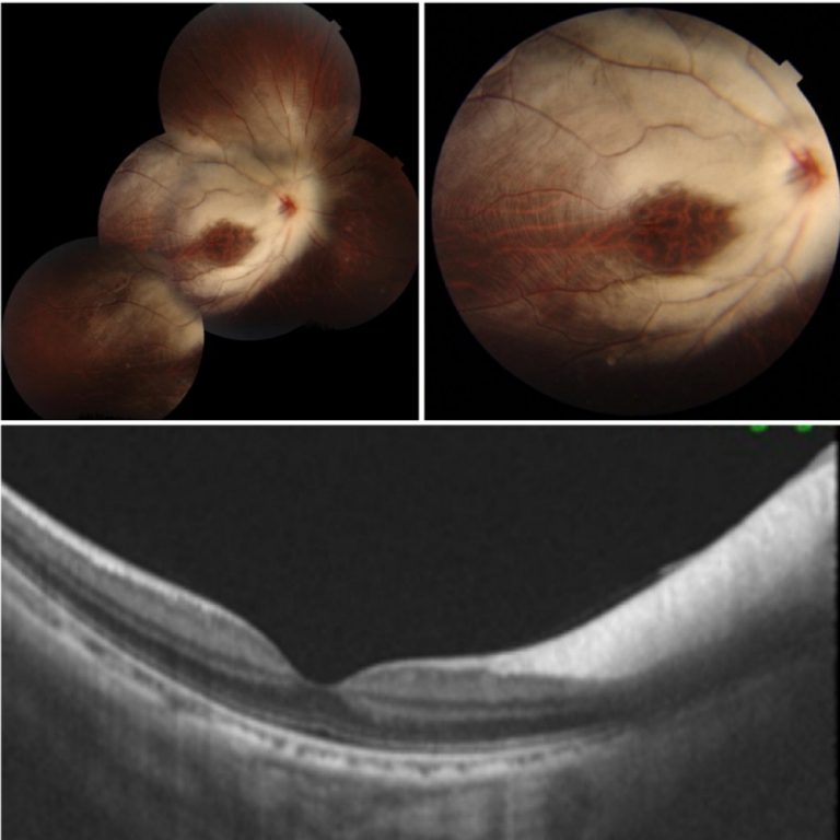
TITLE: Diagnosis : Myelinated nerve fiber ,high myopia with amblyopia.
Case Abstract:-A 9 year old boy came to opd for routine check up .Parents and patient did not notice any complaint.
There was no H/O wearing spectacles. No abnormalities in Antenatal, Natal , postnatal history .Full Term normal delivery and no history of NICU admission was given.
No history of congenital abnormalities.
On examination, Both Eyes ,Anterior segment was within normal limits .Vision in Right Eye was drastically low ,Counting fingers at 1m, Left eye was within normal limits with 6/6 vision.
Based on indirect ophthalmoscopy diagnosis of
Myelinated Nerve fiber with High Myopia with amblyopia was
made. Dilated and undilated refraction gave a retinoscopy value of -15 D in right eye. However, post mydriatic test did not show acceptance of the same neither any improvement of vision .
Patient will be kept on regular follow ups as such patients are prone to neovascularization , branch retinal artery and vein occlusion and vitreous hemorrhage.

DR. PRIYA RASIPURAM CHANDRASEKARAN
Drug induced cortical venous sinus thrombosis (CVST).
A 17 year old girl presented with double vision and headache for 5 days. BCVA was 6/6 N6 in both eyes. Extraocular movements showed left abducent nerve palsy as evidenced by abduction restriction. Anterior segment examination was normal. Fundus examination showed papilledema. History was suggestive of use of oral contraceptive pills (10 mg norethisterone acetate CR) for 4 months for irregular menstruation. MRI brain showed subacute occlusive thrombus seen as hyperintense on T1 sagittal, coronal FLAIR and coronal T2W sequences along the entire superior sagittal sinus and superficial cerebral veins. Coagulation profile for various acquired and inherited thrombophilia, anti-phospholipid antibodies and serum homocysteine were normal. Discontinuation of OC pills and treatment with oral acenocoumarol and subcutaneous heparin caused resolution of symptoms and papilledema. Hence, a proper history and examination followed by timely intervention would lead to favourable outcome and prevent permanent neurological sequelae.


